NIAID Scientists Detail First Structure of a Natural Mammalian Prion

NIAID Now |

NIAID Scientists Detail First Structure of a Natural Mammalian Prion

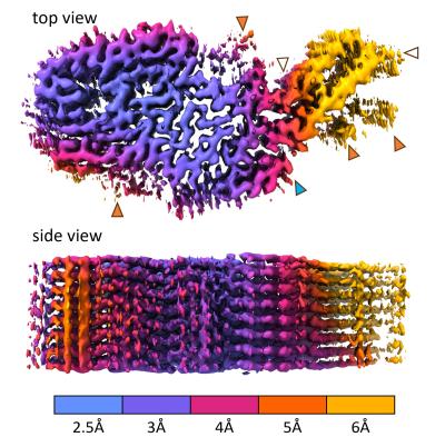
The near-atomic structure of a chronic wasting disease (CWD) prion should help scientists explain how CWD prions spread and become the most naturally infectious of the many mammalian protein aggregation diseases. NIAID scientists revealed the structure in a new study in Acta Neuropathologica. Such detailed knowledge could guide the rational design of vaccines and therapeutics, as well as identify mechanisms that protect humans from CWD pathogens in deer, elk, moose, and reindeer.

Many brain diseases of humans and other mammals involve specific proteins (e.g., prion protein or PrP) gathering into abnormal thread-like structures that grow by sticking to normal versions of the same protein. These threads can also fragment and spread throughout the nervous system and accumulate to deadly levels. For unknown reasons, CWD prions are more naturally contagious than most other protein aggregates and are spreading rampantly among cervid species in North America, Korea and northern Europe. Recalling the bovine spongiform encephalopathy (BSE) or “mad cow disease” epidemic of the mid-1980s and mid-1990s, there are concerns that CWD might similarly be transmissible to humans.

To date, no CWD transmission to humans has been substantiated, and the new CWD structure suggests preliminarily why we might be protected. The structure also reveals multiple differences between CWD and previously determined structures of highly infectious, but experimentally rodent-adapted, PrP-based prions. Differences are even more profound when compared to largely non-transmissible PrP filaments isolated from humans with Gerstmann-Sträussler-Scheinker syndrome, a genetic prion disorder.

PrP-based prion diseases are degenerative, untreatable, and fatal diseases of the central nervous system that occur in people and other mammals. These diseases primarily involve the brain, but also can affect the eyes and other organs. CWD-infected animals shed infectious prions in their feces, urine, and other fluids and body components while alive, and from their carcasses after dying. The prions can remain infectious in the environment for years. 

Scientists at NIAID’s Rocky Mountain Laboratories in Hamilton, Montana, determined the CWD structure from the brain tissue from a naturally infected white-tailed deer. They isolated the prions and froze them in glass-like ice. Then, using electron microscopy techniques, they developed a 3-D electron density map that indicated the detailed shapes of the protein molecules within the prion structure. This involved taking nearly 80,000 video clips of the sample, magnified 105,000 times the original size, at various orientations. They marked prion filaments in the video clips and collected more than 500,000 overlapping sub-images. They isolated about 7,300 of the highest quality sub-images and then used supercomputers to generate a 3-D density map and a molecular model to fit the map.

Vaccine development is among the many research areas where scientists could use high-resolution prion structures to advance their work. The study authors note that previous attempts to develop vaccines against CWD in cervids failed to be protective, and, at least in one case, had the opposite effect. They speculate that one explanation for adverse vaccine effects could be that antibody binding to the sides, rather than the ends of prion fibril surfaces, promotes fragmentation – creating infectious particles rather destroying them. Thus, a strategy to explore with vaccines and small-molecule inhibitors, they say, is to target the tips of prion structures where binding and conversion of prion protein molecules occurs.

The research team is planning to solve other naturally occurring prion structures, hoping to advance its understanding of the molecular basis of prion transmission and disease.

References:

P Alam, F Hoyt, E Artikis, et al. Cryo-EM structure of a natural prion: chronic wasting disease fibrils from deer. Acta Neuropathologica DOI: 10.1007/s00401-024-02813-y (2024).

A Kraus et al. High-resolution structure and strain comparison of infectious mammalian prionsMolecular Cell. DOI: 10.1016/j.molcel.2021.08.011. (2021).

Contact Information

Contact the NIAID Media Team.

301-402-1663
niaidnews@niaid.nih.gov

Search NIAID Blog

The Eyes Have it: A Functional Role for Prion Protein

NIAID Now |

In the early 1980s, scientists identified clumps of abnormal, misfolded prion protein in mammals as the cause of brain-wasting diseases, now called prion diseases. Since that time, they have struggled to answer: What does a normal prion protein do?

The answer, they believe, could help lead them to develop treatments and disease-prevention measures against human prion diseases, such as Creutzfeldt-Jakob disease, fatal familial insomnia and kuru, as well as animal prion diseases, such as scrapie in sheep and chronic wasting disease in cervids.

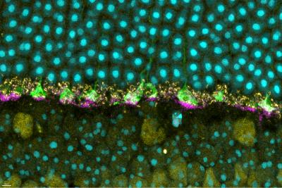
Now, a new study published in iScience from NIAID scientists at Rocky Mountain Laboratories in Hamilton, Montana, and colleagues provides details of how prion protein functions in the retina of mouse eyes, helping them respond to light.

The scientists used mice specially bred without prion protein to compare to wild mice with natural prion protein. Prior studies have suggested that prion protein may have a role in how nerves transmit signals to other nerves at specialized junctions, known as neural synapses. So, knowing that prion protein exists naturally in the eye, the researchers examined mouse retina for a specific neural synapse role.

A key tool the researchers used involved measuring the electroretinographic (ERG) responses – the amount of time it took for the retina in mice to respond to a flash of light. Remember as a kid in school learning about rods and cones in the eye and how they convert light signals to help the brain understand vision? The same is true in mice.

Compared to the wild mice with prion protein, the scientists observed deficiencies in ERG responses for mice without prion protein. The deficiencies affected the normal function of the rods and cones. And – using the ERG data and neural synapse information – they found that the deficiencies originated in the portion of the retina where natural prion protein was most highly concentrated.

Though additional study is needed, the researchers believe the prion protein may act like scaffolding to help cells and elements of the eye, such as rods and cones, to stabilize neural synapses. And they believe prion protein must be present for rods and cones to function normally.

The research team hopes these findings help colleagues who study prion diseases better understand what might occur in humans when natural forms of prion protein are therapeutically removed. New treatment strategies for prion diseases focus on using drugs that remove natural prion protein to eliminate the potential for misfolding and clumping. But researchers do not know whether that could result in unwanted outcomes, such as possibly affecting vision. These findings also could extend to other protein-related neurodegenerative diseases, such as Alzheimer’s (amyloid beta protein) and Parkinson’s diseases (alpha synuclein protein).

Scientists from Duke University and the McLaughlin Research Institute in Great Falls, Montana, collaborated on the study.

Reference: J Striebel, et al. The prion protein is required for normal responses to light stimuli by photoreceptors and bipolar cells. iScience DOI: 10.1016/j.isci.2024.110954 (2024).

Contact Information

Contact the NIAID Media Team.

301-402-1663
niaidnews@niaid.nih.gov

Search NIAID Blog

Household Bleach Inactivates Chronic Wasting Disease Prions

A 5-minute soak in a 40% solution of household bleach decontaminated stainless steel wires coated with chronic wasting disease (CWD) prions, . The scientists used the wires to model knives and saws that hunters and meat processors use when handling deer, elk and moose – all of which are susceptible to CWD.

NIH Study Shows Chronic Wasting Disease Unlikely to Move from Animals to People

A new study of prion diseases, using a human cerebral organoid model, suggests there is a substantial species barrier preventing transmission of chronic wasting disease (CWD) from cervids—deer, elk and moose—to people. The findings, from National Institutes of Health scientists and published in Emerging Infectious Diseases, are consistent with decades of similar research in animal models at the NIH’s National Institute of Allergy and Infectious Diseases (NIAID).

Contact

Submit a Media Request

Contact the NIAID News & Science Writing Branch.

301-402-1663
niaidnews@niaid.nih.gov
All Media Contacts

Novel Study Model Reveals New Understanding of Fatal Familial Insomnia

NIAID Now |

Novel Study Model Reveals New Understanding of Fatal Familial Insomnia
Cerebral Organoids Show NIAID Investigators Disease Characteristics

Fatal familial insomnia (FFI) is a little-known yet horrific disease in which people die from lack of sleep. A protein mutation in the brain prevents sleep, and the body gradually deteriorates. Fortunately, the disease is extremely rare. Fewer than 1,000 people in the United States are estimated to have FFI, according to the NIH’s Genetic and Rare Diseases Information Center. Unfortunately for those with the disease, it can be hereditary – thus the “familial” aspect in the name and importance of developing diagnostic tests and treatments. 

FFI is among a group of unusual neurologic conditions known as prion diseases – those caused by normally harmless prion protein that can malfunction and kill brain cells. Because FFI is found in the brain (suspected origin is the thalamus) studying its spread is not possible until a patient has died. But at that point, valuable disease information is not available because the brain no longer functions. Likewise, studies in rodents and laboratory glassware only have provided limited information.

In a new study published in PLOS Genetics, scientists from the National Institute of Allergy and Infectious Diseases (NIAID) developed a cerebral organoid model to study the exact protein mutation that causes FFI. Human cerebral organoids are small balls of brain cells ranging in size from a poppy seed to a pea; scientists use human skin cells to create organoids. Cerebral organoids have organization, structure, and electrical signaling systems similar to human brain tissue. Because they can survive in a controlled environment for months to years, cerebral organoids also are ideal for studying nervous system diseases over lengthy periods of time.

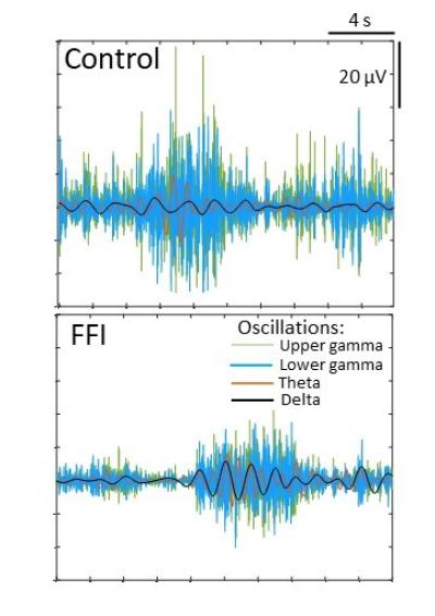
In the new study using the FFI organoids, NIAID scientists working at Rocky Mountain Laboratories in Hamilton, Mont., compared cell functions – primarily in neurons – between the FFI model and organoids without the FFI protein mutation, making several important observations about the mutation’s effect on brain cells.

They believe the abnormalities likely are features of asymptomatic FFI that may lead to disease.

They surprisingly did not observe spontaneous change in shape or spread of the FFI protein to additional prion protein. Such spread typically is a trademark of prion disease – changing prion protein throughout the brain from the normal shape to the malfunctioning folded shape.

“Our findings show that the mutation causes brain cells to dysfunction without the need for misfolding,” the study states. “We could confirm that most changes were caused by the presence of the mutation” rather than interacting with prion protein lacking the mutation.

Further, the group found that neurons in the FFI organoid model were impaired because of damaged mitochondria – which typically produce energy to keep the brain cells healthy. In future studies they hope to establish a relationship between impaired mitochondria function and the mutated FFI prion protein, and whether neurons attempt to stay healthy and avoid harm from the mutated FFI prion protein by switching from mitochondria as an energy source.

They also plan to explore relationships between the mutated FFI prion protein and neurons associated with “wakefulness,” sleep and rapid eye movement in the brain.

Reference:
S Foliaki et al. Altered energy metabolism in Fatal Familial Insomnia cerebral organoids is associated with astrogliosis and neuronal dysfunction. PLOS Genetics DOI: (2023).


Contact Information

Contact the NIAID Media Team.

301-402-1663
niaidnews@niaid.nih.gov

Search NIAID Blog

James A. Carroll, Ph.D.

Education:

Ph.D., 1997, University of Georgia

James A. Carroll, Ph.D.

James A. Carroll, Ph.D.

Associate Scientist


Section or Unit Name
TSE/Prion and Retroviral Pathogenesis Section
First Name
James
Last Name
Carroll
Exclude from directory
Off
Section/Unit: Location
This Researcher/Clinician’s Person Page
Program Description

Dr. Carroll’s primary research is concerned with understanding how neuroinflammation and glial cell activation (astrocytes and microglia) influence prion pathogenesis and neurodegeneration. Prions are infectious misfolded conformers of the cellular GPI-anchored protein PrP and can spread from cell to cell within the brain by seeded-polymerization. This group of proteinopathies can affect humans, cattle, sheep, and cervids and are resistant to many standard decontamination methods. Initially, it was assumed that prion diseases lack an immunological component due to the absence of a prominent antibody or interferon response. However, Dr. Carroll’s research has shown that prion infection elicits a substantial inflammatory response in the CNS and that many inflammatory effectors increase in expression in response to prion infection.

To address the potential impact of microglia in prion disease, Dr. Carroll performed several studies using the potent CSF-R1 inhibitor PLX5622 to reduce microglia in the CNS. These studies indicated that microglia were indispensable to host defense against prion disease. Moreover, his research implicated astrocytes as potentially affecting pathology during disease, where when microglia were absent, the astrocytes were more highly active, expressing numerous disease-related components. This has led to further investigations to assess astrogliosis during prion infection.

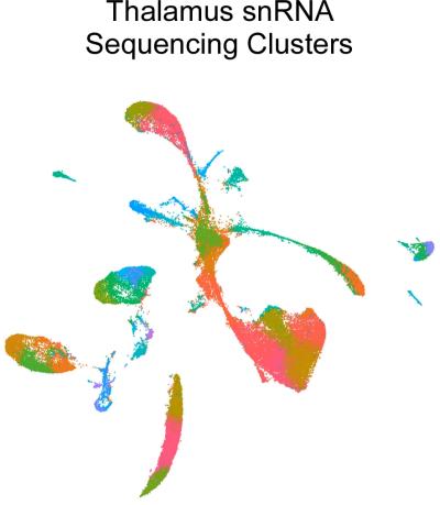
A Uniform Manifold Approximation and Projection (UMAP) of single nuclei RNA (snRNA) sequencing analysis of uninfected and prion-infected mouse thalamus depicting 43 transcriptional clusters from >69,000 nuclei examined.

A Uniform Manifold Approximation and Projection (UMAP) of single nuclei RNA (snRNA) sequencing analysis of uninfected and prion-infected mouse thalamus depicting 43 transcriptional clusters from >69,000 nuclei examined.

Credit: NIAID

Using high-throughput deep sequencing of RNA transcripts in longitudinal studies, Dr. Carroll has identified numerous differentially expressed genes in the CNS during prion infection. These investigations have yielded compelling results and suggest that microglia in the prion-infected brain assume an alternative phenotype that is distinct from those seen in other brain disorders. From these RNA-seq studies, it was determined that reactive astrocytes assume an expression signature that is not reliant on the canonical signals described in other neuroinflammatory models. Furthermore, this prion-specific reactive astrocyte expression signature is exacerbated when microglia are reduced in the CNS.

Dr. Carroll has begun to analyze the individual cellular changes in the brain using single nuclei RNA (snRNA) sequencing to better understand the relevant changes in the cell populations in the complex milieu of the CNS during infection. Thus far, the research has focused on gene expression changes in the thalamus at pre- and early-clinical times. The thalamus is affected early during prion infection in rodent models, and thalamic pathology is a key feature in human forms of prion disease.

A new aspect of Dr. Carroll’s research is a collaboration with Dr. Cathryn Haigh (Chief, Prion Cell Biology Unit, NIAID) to study Lyme Neuroborreliosis (LNB). Lyme disease, a global public health concern, is the most common tick-borne disease in North America and Eurasia, with an estimated 14% of the world’s population having become infected. Reported cases of Lyme disease in the U.S. have been on the rise for many years, with over 62,000 confirmed cases reported in 2022, making it the leading reportable arthropod-borne infectious disease. The Centers for Disease Control estimates that the disease is underreported, and the true incidence of Lyme disease in the U.S. is approaching 500,000 cases annually. Lyme disease, caused by bacterial spirochetes of the genus Borrelia, is a multi-systemic disorder affecting the skin, heart, central nervous system, and joints.

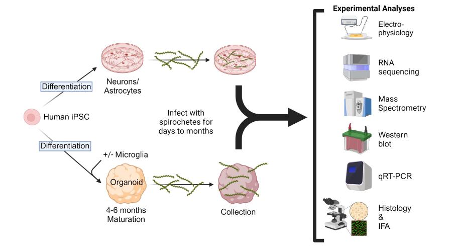
To address the need for additional models of LNB and to better understand the responses of the human CNS when exposed to Borrelia, this collaboration has developed two in vitro model systems. The first uses human cerebral organoids differentiated from human induced pluripotent stem cells (iPSCs) as an in vitro tissue model. The second uses iPSCs for differentiation into specific neuronal subtypes, astrocytes, and microglia-like cells for study. Exploiting these human-derived model systems, we are assessing responses to Borrelia infection that are stimulated in isolated cells from specific responses that only occur in these cells when they are part of an integrated organoid network. This project incorporates several cutting-edge technologies, including organoid development, bulk and single-cell RNA sequencing, metabolomics, and lipidomics.

Experimental design and strategy to address potential responses of human cerebral organoids, astrocytes, and neurons after exposure to infectious Borrelia species that cause Lyme Neuroborreliosis.

Experimental design and strategy to address potential responses of human cerebral organoids, astrocytes, and neurons after exposure to infectious Borrelia species that cause Lyme Neuroborreliosis.

Credit: NIAID
Selected Publications

Carroll JA, Striebel JF, Baune C, Chesebro B, Race B. CD11c is not required by microglia to convey neuroprotection after prion infection. PLoS One. 2023 Nov 1;18(11):e0293301.

Carroll JA, Race B, Williams K, Striebel JF, Chesebro B. Innate immune responses after stimulation with Toll-like receptor agonists in ex vivo microglial cultures and an in vivo model using mice with reduced microglia. J Neuroinflammation. 2021 Sep 6;18(1):194.

Carroll JA, Foliaki ST, Haigh CL. A 3D cell culture approach for studying neuroinflammation. J Neurosci Methods. 2021 Jul 1;358:109201.

Carroll JA, Race B, Williams K, Striebel J, Chesebro B. RNA-seq and network analysis reveal unique glial gene expression signatures during prion infection. Mol Brain. 2020 May 7;13(1):71.

Carroll JA, Race B, Williams K, Striebel J, Chesebro B. Microglia Are Critical in Host Defense against Prion Disease. J Virol. 2018 Jul 17;92(15):e00549-18.

Carroll J.A., J.F. Striebel, A. Rangel, T. Woods, K. Phillips, K.E. Peterson, B. Race, and B. Chesebro. 2016. Prion strain differences in accumulation of PrPSc on neurons and glia are associated with similar expression profiles of neuroinflammatory genes: comparison of three prion strains. PLoS Path. Apr 5;12(4):e1005551.

Visit PubMed for a complete publication list.

Additional Information

Awards

  • 1990 Graduated Cum Laude from Clemson University.            
  • 1993 and 1994 Recipient: Outstanding performance in research and teaching merit award from the Graduate School of the University of Georgia.
  • 1997-2002 Recipient: Intramural Research Training Award (IRTA), NIAID, NIH.    
  • 2001 Recipient: NIAID Richard Asofsky Special Achievement Award in Equal Employment Opportunity in recognition of participation in the B.R.A.S.S. program.
  • 2011 Recipient: the James H. Nakano Citation from the Centers for Disease Control for outstanding scientific article Gilmore et al. 2010. PNAS. 107(16):7515-7520.
  • 2011 Recipient: the Charles C. Shepard Science Award, the highest CDC award for excellence in science, for an outstanding scientific article published in 2010 (Gilmore et al. PNAS. 107(16):7515-7520).
  • 2019 Recipient: National Institutes of Health, NIAID 10 Years of Service award.
  • 2021 Recipient: Honorific title of Associate Scientist in recognition of exceptional achievements as a Staff Scientist in the NIAID Division of Intramural Research.
     
Major Areas of Research
  • Neuroinflammation during preclinical and clinical prion infection
  • Influence of microglia and neurotoxic astrocytes on prion pathogenesis
  • Alterations in cell populations and gene expression in the central nervous system and retina after prion infection
  • Modeling Neuroborreliosis in human-derived neurons, astrocytes, and organoids

NIAID Study Reveals, Compares Prion Strains

NIAID Now |

Prions are infectious protein pathogens that cause fatal neurodegenerative diseases in mammals. For decades, scientists have wondered how different strains of prions can propagate when they do not carry their own genes with them as they move from host to host. A new study from NIAID researchers and colleagues at Case Western Reserve University in Cleveland reveals – in near-atomic detail – how differences in the folding of the primary protein of prions (PrP) can help determine the distinct characteristics of prion strains.

The research teams are pursuing studies of prion structure to aid in the design and screening of treatments that might slow or prevent the spread of these deadly diseases. Prion diseases are caused by the corruption of the normal form of PrP that is made by all mammals. Although poorly understood, normal PrP molecules spend most of their time as individual units that play roles in multiple physiological functions.

Rarely, and for unknown reasons, normal PrP molecules can spontaneously refold into highly structured assemblies that cause brain disease. This spontaneous process seems to be responsible for the most common form of prion disease in humans, sporadic Creutzfeldt-Jakob disease. However, once formed, corrupted PrP aggregates, or prions, can be highly infectious if inadvertently transferred from one person to another by invasive medical procedures. In other mammals with poorer basic hygiene, such as livestock and wildlife, prion infections such as scrapie, chronic wasting disease, or mad cow disease can spread by more casual contact and contaminate the environment.

Multiple strains of prions have been identified in all these host species, with each giving a distinctive array of clinical presentations, molecular features, patterns of damage in the brain, and transmissibility to other species. Thus, deciphering the basis of prion strain diversity is important in understanding the risks posed by prions to which humans or animals are exposed.

Prions can reproduce in hosts a billion times or more during infection. They do so without having their own infection-specific genes because all PrP molecules are encoded by the same host gene. For this reason, scientists cannot easily track prions like they can viruses and bacteria.

The new study, published in Nature Communications, comes from the same researchers who in 2021 published the first high-resolution structure of an infectious prion protein. That work solved the structure of a specific hamster-adapted strain of scrapie, a prion disease that occurs naturally in sheep and goats. The initial study also reported lower resolution images of a mouse-adapted form of scrapie that already indicated strain-dependent differences in overall shape. 

The latest study uses cryo-electron microscopy to show the structure of this mouse-adapted second prion strain in much greater detail. The strain was depicted at a resolution of about 3 angstroms in size (1 angstrom is equal to one hundred-millionth of a centimeter). Comparing these rodent prion strains reveals that, although they share some key structural similarities, their details greatly differ overall.

Importantly, both strains are pancake-like layers of PrP molecules with a ladder-like protein substructure. Prior to the 2021 study, scientists only had educated guesses to guide their work regarding prion characteristics.

“The structural basis for how prions replicate as deadly pathogens, and to do so consistently as different strains to cause distinct diseases, has long been a major mystery in our field of research,” Dr. Byron Caughey, the senior NIAID scientist on the study, said. “Now we have a much clearer idea of how this works – at least for these first two strains that we have solved so far.”

References:
F Hoyt et al. Cryo-EM structure of anchorless RML prion reveals variations in shared motifs between distinct strains. Nature Communications. DOI: https://doi.org/10.1038/s41467-022-30458-6 (2022).

A Kraus et al. High-resolution structure and strain comparison of infectious mammalian prions. Molecular Cell. DOI: 10.1016/j.molcel.2021.08.011. (2021).

Contact Information

Contact the NIAID Media Team.

301-402-1663
niaidnews@niaid.nih.gov

Search NIAID Blog

Karin Peterson, Ph.D.

Education:

Ph.D., 1998, University of Missouri Medical School

Karin Peterson, Ph.D.

Suzette A. Priola, Ph.D.

Education:

Ph.D., 1990, University of California, Los Angeles

Photo of Suzette A. Priola, Ph.D.