Natural History Study of GATA2 Deficiency and Related Disorders

The purpose of this study is to improve understanding of GATA2 deficiency so there can be better diagnostic tests and treatments in the future.

A Single-arm, Dose-escalation Trial of Long-acting Recombinant Human IL-7 (NT-I7, Efineptakin Alfa) for Idiopathic CD4 Lymphopenia

The purpose of this study is to test a new drug (NT-17) in people with idiopathic CD4 lymphopenia (ICL) which can increase the number of CD4 T cells.

Contact Information

Office/Contact: For more information at the NIH Clinical Center contact Office of Patient Recruitment (OPR)
Phone: 800-411-1222
TTY: TTY dial 711
Email: ccopr@nih.gov
 

Novel Genetic Disorders of the Immune System

The purpose of this study is to evaluate people with certain types of immune system disorders.

NIAID Scientists Detail First Structure of a Natural Mammalian Prion

NIAID Now |

NIAID Scientists Detail First Structure of a Natural Mammalian Prion

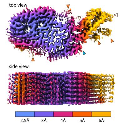
The near-atomic structure of a chronic wasting disease (CWD) prion should help scientists explain how CWD prions spread and become the most naturally infectious of the many mammalian protein aggregation diseases. NIAID scientists revealed the structure in a new study in Acta Neuropathologica. Such detailed knowledge could guide the rational design of vaccines and therapeutics, as well as identify mechanisms that protect humans from CWD pathogens in deer, elk, moose, and reindeer.

Many brain diseases of humans and other mammals involve specific proteins (e.g., prion protein or PrP) gathering into abnormal thread-like structures that grow by sticking to normal versions of the same protein. These threads can also fragment and spread throughout the nervous system and accumulate to deadly levels. For unknown reasons, CWD prions are more naturally contagious than most other protein aggregates and are spreading rampantly among cervid species in North America, Korea and northern Europe. Recalling the bovine spongiform encephalopathy (BSE) or “mad cow disease” epidemic of the mid-1980s and mid-1990s, there are concerns that CWD might similarly be transmissible to humans.

To date, no CWD transmission to humans has been substantiated, and the new CWD structure suggests preliminarily why we might be protected. The structure also reveals multiple differences between CWD and previously determined structures of highly infectious, but experimentally rodent-adapted, PrP-based prions. Differences are even more profound when compared to largely non-transmissible PrP filaments isolated from humans with Gerstmann-Sträussler-Scheinker syndrome, a genetic prion disorder.

PrP-based prion diseases are degenerative, untreatable, and fatal diseases of the central nervous system that occur in people and other mammals. These diseases primarily involve the brain, but also can affect the eyes and other organs. CWD-infected animals shed infectious prions in their feces, urine, and other fluids and body components while alive, and from their carcasses after dying. The prions can remain infectious in the environment for years. 

Scientists at NIAID’s Rocky Mountain Laboratories in Hamilton, Montana, determined the CWD structure from the brain tissue from a naturally infected white-tailed deer. They isolated the prions and froze them in glass-like ice. Then, using electron microscopy techniques, they developed a 3-D electron density map that indicated the detailed shapes of the protein molecules within the prion structure. This involved taking nearly 80,000 video clips of the sample, magnified 105,000 times the original size, at various orientations. They marked prion filaments in the video clips and collected more than 500,000 overlapping sub-images. They isolated about 7,300 of the highest quality sub-images and then used supercomputers to generate a 3-D density map and a molecular model to fit the map.

Vaccine development is among the many research areas where scientists could use high-resolution prion structures to advance their work. The study authors note that previous attempts to develop vaccines against CWD in cervids failed to be protective, and, at least in one case, had the opposite effect. They speculate that one explanation for adverse vaccine effects could be that antibody binding to the sides, rather than the ends of prion fibril surfaces, promotes fragmentation – creating infectious particles rather destroying them. Thus, a strategy to explore with vaccines and small-molecule inhibitors, they say, is to target the tips of prion structures where binding and conversion of prion protein molecules occurs.

The research team is planning to solve other naturally occurring prion structures, hoping to advance its understanding of the molecular basis of prion transmission and disease.

References:

P Alam, F Hoyt, E Artikis, et al. Cryo-EM structure of a natural prion: chronic wasting disease fibrils from deer. Acta Neuropathologica DOI: 10.1007/s00401-024-02813-y (2024).

A Kraus et al. High-resolution structure and strain comparison of infectious mammalian prionsMolecular Cell. DOI: 10.1016/j.molcel.2021.08.011. (2021).

Contact Information

Contact the NIAID Media Team.

301-402-1663
niaidnews@niaid.nih.gov

Search NIAID Blog

NIAID Enables Approval of Novel Anti-TB Drug

Sequencing of Congo Mpox Reports Highlights New Transmission Patterns in Country

NIAID Now |

Country-led genetic analysis of samples collected through the Republic of Congo (RoC) epidemiologic surveillance system in early 2024 showed that mpox was affecting people in parts of the country where it has not been historically reported, and point to increases in human-to-human transmission across the border with the neighboring Democratic Republic of the Congo (DRC), where a large outbreak was declared a public health emergency of international concern in August of the same year. The analysis was conducted by the RoC Laboratoire National de Santé Publique (LNSP) in Brazzaville with support and scientific partnership from NIAID and was published in The Lancet

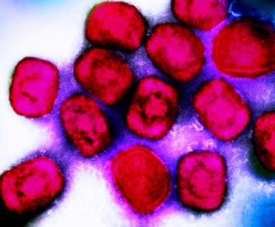
There are two known types or “clades” of monkeypox virus (MPXV), which causes mpox clinical disease. Clade I is endemic in Central Africa and can cause severe illness. Clade II, endemic in West Africa, caused the global mpox outbreak that began in 2022 and tends to result in milder illness. Each clade has two known subtypes referred to as “a” and “b.” Clade Ia has been identified in RoC and DRC intermittently for decades and Clade Ib was first identified during the active DRC outbreak. Mpox is a zoonotic disease, meaning it can be spread between animals and people. MPXV has been detected in rodents that live in areas historically affected by mpox. 

Genetic sequencing of MPXV can help determine the transmission dynamics and guide the public health response to mpox, but until recently most sequencing of MPXV was done outside of affected countries like RoC, requiring costly sample transport and delaying decision-making by local health authorities. 

To better understand whether mpox in RoC was driven by spillover from local animal hosts or cross-border human-to-human transmission from DRC, a team led by the RoC LNSP analyzed 31 samples of laboratory-confirmed MPXV collected through the country’s routine epidemiologic surveillance system between January and April of 2024. Using new in-country sequencing technology, the team determined that there were diverse circulating strains of MPXV in the country, all of the Clade Ia subtype, and some showed up to 99.9% genetic similarity to MPXV sequenced from the DRC. Moreover, MPXV samples came from provinces without historical reports of mpox. 

According to the authors, the diversity of identified stains suggest MPXV has been introduced to the human population in RoC through multiple distinct events, which could be a combination of direct zoonotic transmission from local animals as well as human-to-human transmission within and across the country’s borders. They state that current epidemiological data are insufficient to definitively confirm the directionality of MPXV transmission and that further epidemiological research is needed to understand local transmission patterns and inform the public health response in RoC. Finally, they highlight that while only 31 samples met criteria for analysis in the study, it is likely these cases represent only a fraction of the RoC mpox burden at the time of collection.

This research informed the RoC’s decision to declare a national mpox epidemic in April 2024. It is part of a longstanding scientific collaboration between NIAID’s Rocky Mountain Laboratories and the Congolese government. The U.S. Embassy in RoC, the U.S. Agency for International Development, the U.S. Centers for Disease Control and Prevention, and the World Health Organization also provided technical and laboratory support for this study. 

Learn more about NIAID’s mpox research priorities. Play a video of NIAID Director Jeanne Marrazzo discussing these priorities. 

Reference:

CK Yinda, et al. Genetic sequencing analysis of monkeypox virus clade I in Republic of the Congo: a cross-sectional, descriptive study. The Lancet DOI: 10.1016/S0140-6736(24)02188-3 (2024)

Contact Information

Contact the NIAID Media Team.

301-402-1663
niaidnews@niaid.nih.gov

Search NIAID Blog

Kidney Transplantation Between Donors and Recipients with HIV Is Safe

Kidney transplantation from deceased donors with HIV to recipients with HIV was safe and comparable to kidney transplantation from donors without HIV.

Contact

Submit a Media Request

Contact the NIAID News & Science Writing Branch.

301-402-1663
niaidnews@niaid.nih.gov
All Media Contacts

NIAID Mouse Study Shows Existing Drug Could Limit COVID-19 Lung Damage

NIAID Now |

A drug that blocks danger signals that can lead to harmful inflammation could help reduce COVID-19 lung damage, a new study from NIAID scientists and colleagues has found. Scientists from NIAID’s Rocky Mountain Laboratories in Hamilton, Montana, and the University of Utah completed the project, published online in JCI Insights.

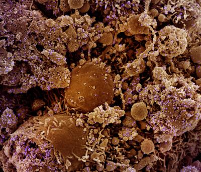
Though they completed the study in laboratory mice modified to model COVID-19 in people, the scientists think their findings are important enough to pursue further studies of the drug, FPS-ZM1, to determine dosing and timing strategies for possible human clinical trials. FPS-ZM1 is an immune modulatory therapeutic – the drug is designed to prevent a specific immune system response from occurring. The investigational therapy has been evaluated in preclinical studies to treat conditions such as diabetes, lung injury and stroke. In their study, the scientists used FPS-ZM1 to block the “receptor for advanced glycation end products” (RAGE), which senses danger signals and can generate inflammation and coagulation known to damage the lungs of COVID-19 patients. 

Therapeutic treatment with FPS-ZM1 during the study improved survival in mice infected with SARS-CoV-2, the virus that causes COVID-19. Further, FPS-ZM1 specifically reduced damage to the lung vasculature, an important system for circulating blood through the lungs that becomes damaged during SARS-CoV-2 infection. FPS-ZM1 also has shown in other rodent studies that it can protect against injury in disease models of brain injury, sepsis, asthma, diabetes, acute lung injury and ischemic/reperfusion (organ damage due to blood flow).

The study also identified two distinct phases of COVID-19 disease development in the mice. The scientists want to further explore those phases as potential guides for treatment strategies. For example, FPS-ZM1 limited specific types of inflammation and tissue damage, so it would likely be most effective if administered during the intermediate to later stages of SARS-CoV-2 infection, whereas antiviral treatment may be most effective when given early following infection.

Reference: F Jessop, et al. Impairing RAGE signaling promotes survival and limits disease pathogenesis following SARS-CoV-2 infection. JCI Insights DOI: https://doi.org/10.1172/jci.insight.155896. (2022).
 

Contact Information

Contact the NIAID Media Team.

301-402-1663
niaidnews@niaid.nih.gov

Search NIAID Blog

COVID-19 Respiratory Treatment Effective in Encephalitis Study

NIAID Now |

COVID-19 Respiratory Treatment Effective in Encephalitis Study

Molnupiravir Reduced Viral Brain Disease in Mice

NIAID research into finding broad uses for existing drug treatments has a potential new success story: Molnupiravir, a relatively new antiviral developed to treat respiratory diseases – such as COVID-19 – reduced brain swelling in study mice infected with a pathogen dangerous to children, La Crosse virus (LACV).

The new study, from NIAID scientists and colleagues at the University of North Carolina and Emory University, is published in PLOS Pathogens. LACV, which is spread by mosquitos, can cause brain inflammation in children. LACV was first isolated in the early 1960s near La Crosse, Wisconsin. Since then, LACV encephalitis cases have been found in more than 20 states, mostly in the basins of the Mississippi and Ohio rivers and throughout the Appalachian Mountains.

Most LACV infections in people are mild, but the virus sometimes – particularly in children – enters the brain, infects neurons and causes disease that can result in learning and memory difficulties, paralysis, seizures and death. Between 30 and 90 cases of severe LACV – those that affect the central nervous system (CNS) – are reported each year, though the Centers for Disease Control and Prevention believes many mild cases occur but are not diagnosed.

The study used a new strategy to test three antiviral drugs – N4-Hydroxycytidine (NHC, the active metabolite of the prodrug molnupiravir), ribavirin and favipiravir– for treatment against LACV infection. The scientists chose LACV because it broadly represents several RNA viruses that cause disease in the CNS, including Jamestown Canyon and Cache Valley viruses – which also were part of the study – and rabies, polio, West Nile, Nipah and several other viruses not part of the study.

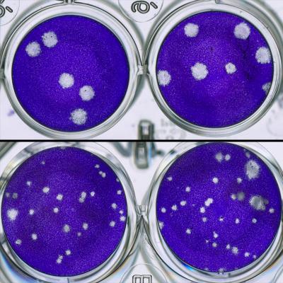
The three antiviral drugs were tested in a cell culture system to examine an antiviral strategy called lethal mutagenesis. This approach increases the number of errors in the viral genome that RNA viruses make when they replicate, weakening the resulting viruses. By incorporating the drug, more errors are induced in the viral genome and more weakened viruses emerge, providing the host an opportunity to recover.

Ribavirin and favipiravir used in cell treatment studies did not produce potent enough results to justify testing in mice. The NHC prodrug molnupiravir, however, was used in two different mouse study models. Oral treatment with molnupiravir reduced brain disease in mice by 32% when LACV infection was started by an injection in the abdomen, and by 23% when the infection was started in the nose, offering easy access to the brain.

Also noteworthy: The researchers tested NHC against LACV and found it effective in the cell and mouse models, as well as in cell models using Jamestown Canyon and Cache Valley viruses. This showed that the drug treatment strategy could be successful against viruses related to LACV and supports the idea that this strategy could be used against a broader group of encephalitic RNA viruses.

The researchers say more study is needed to see how these drugs counter RNA viruses, particularly to determine whether injecting the drug directly into cerebrospinal fluid would provide better results and possibly reduce adverse side effects.

Reference:

D Ojha et alN4-Hydroxycytidine/Molnupiravir Inhibits RNA Virus-Induced Encephalitis by Producing Less Fit Mutated VirusesPLOS Pathogens DOI: 10.1371/journal.ppat.1012574 (2024).

Contact Information

Contact the NIAID Media Team.

301-402-1663
niaidnews@niaid.nih.gov

Search NIAID Blog

Tiny Nanoparticles Could Be A Big Jump for Flu Vaccines