Fernanda D. Young, M.D.

Specialty(s): Allergy and Immunology, Internal Medicine, Pediatrics
Provides direct clinical care to patients at NIH Clinical Center

Education:

M.D., University of Washington School of Medicine, Seattle, WA

Languages Spoken: Spanish
Fern Young Headshot

Assessment of PrEP Eligibility Tool Among the General Population in Uganda

NIAID Now |

To reduce the spread of HIV infection, the World Health Organization (WHO) recommends pre-exposure prophylaxis (PrEP) in populations with an annual HIV infection incidence greater than 3%. Given that the annual HIV incidence in Uganda is estimated at 0.40% (0.46% among females and 0.35% among males), there are people who may have a higher HIV risk and may benefit from PrEP but are not targeted for services. To further reduce the spread of HIV in Uganda, researchers used results from three survey rounds of HIV-negative participants within the Rakai Community Cohort Study (RCCS) to estimate the prevalence of high-risk individuals eligible for PrEP within the general population and the incidence of HIV infection associated with eligibility. 

In this study, a subset of questions from Uganda’s PrEP eligibility tool that are routinely asked within the RCCS surveys were used to determine PrEP eligibility. Eligibility was defined as reporting at least one of the following HIV risk behaviors in the past 12 months: sexual intercourse with more than one partner of unknown HIV status; nonmarital sex act without a condom; sex engagement in exchange for money, goods, or services; or experiencing genital ulcers. HIV incidence was estimated by analyzing seroconversion from HIV-negative to HIV-positive in participants who contributed to at least two of the survey rounds.

Overall, 29% of participants in the analysis met the eligibility criteria. Of these, 22% reported one HIV risk, 6% reported two HIV risks, and 1% reported three HIV risks. The results showed that PrEP eligible participants had twice the risk of acquiring HIV than their non-eligible counterparts. Furthermore, risk increased threefold in uncircumcised males but not circumcised males. Additionally, men who reported higher prevalence of risky behaviors had lower increase in HIV incidence compared to women, likely due to circumcision status and higher antiretroviral therapy coverage in HIV-infected females, leading to a decrease in transmission to men. The findings of this study support the use of PrEP eligibility screening in general populations with HIV incidence lower than 3% to reduce HIV acquisition even further in these populations.

Reference: Ssempijja et al. High Rates of Pre-exposure Prophylaxis Eligibility and Associated HIV Incidence in a Population With a Generalized HIV Epidemic in Rakai, Uganda, JAIDS Journal of Acquired Immune Deficiency Syndromes, July 1, 2022 - Volume 90 - Issue 3 - p 291-299.

Contact Information

Contact the NIAID Media Team.

301-402-1663
niaidnews@niaid.nih.gov

Search NIAID Blog

First FDA-Approved Treatment for Eosinophilic Esophagitis Has Roots in NIAID-Funded Research

NIAID Now |

The first Food and Drug Administration approval of a treatment for eosinophilic esophagitis, announced in May 2022, marked a vital achievement not only for people with the disease, but also for scientists including a NIAID grantee whose research helped lay a foundation for this milestone.

Eosinophilic esophagitis, or EoE, is a chronic disease characterized by an overabundance of a specific type of white blood cell, an eosinophil, in the esophagus. The disease is driven by allergic inflammation due to food. This inflammation damages the esophagus and prevents it from working properly. For people with EoE, swallowing even small amounts of food can be a painful and worrisome choking experience. EoE is the most common reason people must go to emergency departments for a healthcare provider to remove food stuck in their esophagus. People with EoE are often left to contend with the frustration and anxiety of a sometimes-lengthy list of foods to avoid, a poor quality of life, and a higher risk of depression. In severe cases, a feeding tube may be the only option to ensure proper caloric intake and adequate nutrition. About 160,000 people in the United States are living with EoE.

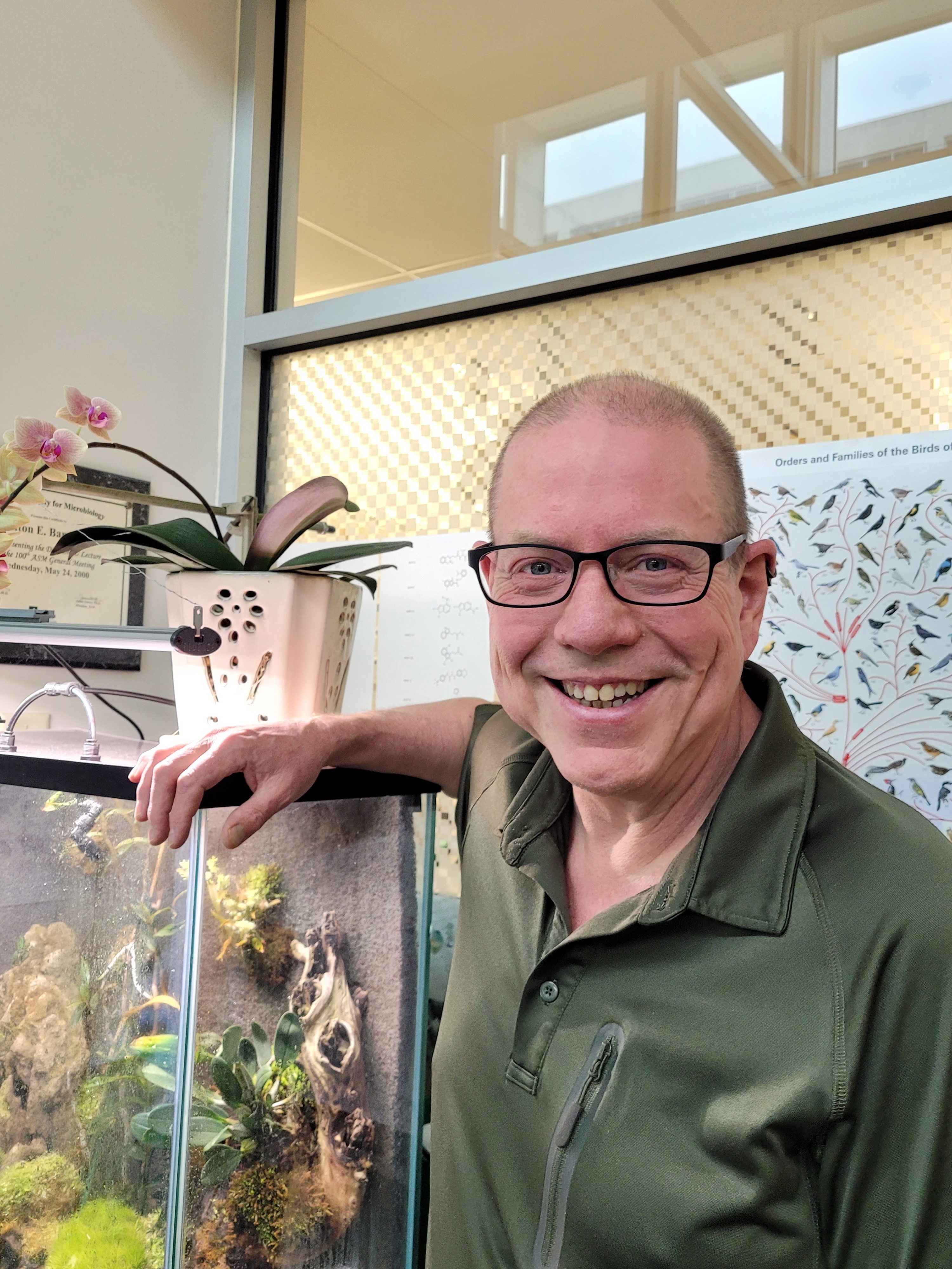
NIAID funding enabled Marc E. Rothenberg, M.D., Ph.D., at Cincinnati Children’s in Ohio to conduct basic and preclinical research starting in 1999 that uncovered the molecular cause of EoE. This finding suggested the type of drug needed to treat the disorder.

Before 2001, some in the medical community thought EoE was a form of acid reflux disease. Dr. Rothenberg’s lab published a paper that year establishing that EoE is actually an allergic disorder. The paper showed that mice exposed to an inhaled respiratory allergen developed EoE. This helped focus subsequent studies of EoE on the role of allergic inflammation.

A few years later, two seminal papers from Dr. Rothenberg’s lab identified the molecular changes taking place in the esophagus of people with EoE and showed that a cell-signaling molecule called IL-13 was responsible for many of those changes. IL-13 and another cell-signaling molecule, IL-4, together drive allergic inflammation in many diseases.

The first of these two key papers reported the results of an analysis of gene expression in cells on the lining of the esophagus in people with and without EoE. The study identified 574 genes that were copied into RNA “transcripts”—the instructions for making proteins—in greater or smaller numbers in people with EoE than in healthy people. This EoE transcript signature, or transcriptome, served as a key reference for subsequent studies of the disorder.

The second paper demonstrated in 2007 that many of the EoE-associated genes identified in the earlier paper are directly activated by IL-13 in cells lining the esophagus, implicating this molecule as a major regulator of the biological pathways involved in EoE. This finding suggested that IL-13-blocking drugs might effectively treat the disease. The study further showed that 98% of the EoE transcriptome reverted to normal levels of gene expression in people whose EoE was successfully treated with a class of steroid hormones called glucocorticoids. This indicated that the EoE transcriptome changes in response to changes in signs and symptoms of the disease. Dr. Rothenberg and colleagues therefore proposed using the EoE transcriptome to monitor the efficacy and mechanism of action of IL-13-blocking drugs at the molecular level.

Several years later, Dr. Rothenberg designed and led the first clinical trial to test the efficacy of an anti-IL-13 monoclonal antibody for treating EoE. The investigators found that the antibody lowered levels of eosinophils in the esophagus and returned the expression of 29 key genes in the EoE transcriptome to normal levels in most treated participants. This and related molecular analyses from the trial, funded by Novartis Pharma AG of Basel, Switzerland, further supported Dr. Rothenberg’s theory that EoE was driven by IL-13.

These findings contributed to a body of foundational EoE research developed by a multitude of scientists and physicians. This scientific foundation, among many other factors, led Regeneron Pharmaceuticals Inc. of Tarrytown, New York, and Sanofi of Paris to begin testing one of their drugs for the treatment of EoE. The drug, a monoclonal antibody called dupilumab, works by blocking both IL-13 and IL-4.

Ultimately, Regeneron and Sanofi conducted a Phase 3 clinical trial showing dupilumab substantially improved the signs and symptoms of EoE compared to a placebo. Based on these results, FDA approved dupilumab for the treatment of the disease on May 20, 2022, making it the first medicine specifically indicated to treat EoE in the United States.

NIAID-funded basic and translational research continues to contribute to the development of preventive and therapeutic strategies for dozens of allergic, immunologic, and infectious diseases.

References:

A Mishra, et al. An etiological role for aeroallergens and eosinophils in experimental esophagitis. The Journal of Clinical Investigation DOI: 10.1172/JCI10224 (2001).

C Blanchard, et al. Eotaxin-3 and a uniquely conserved gene-expression profile in eosinophilic esophagitis. The Journal of Clinical Investigation DOI: 10.1172/JCI26679 (2006).

C Blanchard, et al. IL-13 involvement in eosinophilic esophagitis: Transcriptome analysis and reversibility with glucocorticoids. Journal of Allergy and Clinical Immunology DOI: 10.1016/j.jaci.2007.10.024 (2007).

ME Rothenberg, et al. Intravenous anti–IL-13 mAb QAX576 for the treatment of eosinophilic esophagitis. Journal of Allergy and Clinical Immunology DOI: 10.1016/j.jaci.2014.07.049 (2015).

Contact Information

Contact the NIAID Media Team.

301-402-1663
niaidnews@niaid.nih.gov

Search NIAID Blog

John Misasi, M.D.

Specialty(s): Infectious Disease, Pediatrics
Provides direct clinical care to patients at NIH Clinical Center

Education:

M.D., 2002, SUNY Upstate Medical University, Syracuse, NY

B.S., 1996, Boston University, MA

NIH Continues Investment in HIV Prevention, Therapy and Cure with $34 Million Award

NIAID Now |

The National Institutes of Health has awarded approximately $34 million annually over the next five years to fund six independent Centers for HIV Structural Biology. The National Institute of Allergy and Infectious Diseases (NIAID) committed approximately $30 million in funding, with an additional $4 million from the NIH Office of AIDS Research.

The Centers program, established in 2007 by the National Institute of General Medical Sciences (NIGMS), was transitioned to NIAID in 2019 as part of the transfer of the NIGMS HIV/AIDS portfolio to the NIAID Division of AIDS (DAIDS). Last year, NIAID solicited applications to RFA AI-21-030: Centers for HIV Structural Biology using the U54 specialized center funding mechanism. The U54 mechanism was utilized to allow enhanced NIAID program oversight and encourage the involvement of NIH intramural researchers. The new Centers largely maintained the structure of the existing Centers while refocusing on research topics that support NIAID priorities of HIV prevention, therapy and cure. In addition, a new Developmental Core was added to foster mentoring and training of young scientists in HIV research.

The Center researchers integrate techniques from structural biology, biochemistry and cell biology to capture in unprecedented detail the three-dimensional structures of HIV proteins and nucleic acids and their interactions with cellular components. This information provides an entirely new perspective on HIV infection and immune control that helps elucidate how the different components interact and reveal new approaches for disrupting those interactions, potentially leading to new targets for HIV therapies and preventative vaccines. In total over 300 scientists from dozens of institutions in the US and abroad will participate in the studies put forward by the Centers. 

The six applications selected for funding include research across the HIV life cycle, aiming, for example, to elucidate the mechanism the HIV envelope protein uses to enter a target cell, the interactions of virus structures with host proteins that either facilitate or antagonize infection, and the way that the HIV RNA genome folds into complex assemblies with other viral components before being packaged into newly formed virions.

The following awardees will receive the indicated funds annually for five years: 

Duke University

Duke Center for HIV Structural Biology (DCHSB)

Director: Priyamvada Acharya

Award: $5.5 million


Seattle Children’s Hospital

Behavior of HIV in Viral Environments (B-HIVE)

Co-Directors: Bruce Torbett, Stefan Sarafianos

Award: $6.3 million 


University of California, San Francisco

HIV Accessory and Regulatory Complexes (HARC)

Director: Nevan Krogan

Award: $5.7 million


University of Michigan

Center for Structural Biology of HIV RNA (CRNA)

Director: Alice Telesnitsky

Award: $5.5 million


University of Pittsburgh

Pittsburgh Center for HIV Protein Interactions (PCHPI)

Directors: Angela Gronenborn, Tatyana Polenova

Award: $5 million


University of Utah

Center for the Structural Biology of HIV Infection, Restriction, and Viral Dynamics (CHEETAH)

Director: Wesley Sundquist

Award: $5.5 million


Contact Information

Contact the NIAID Media Team.

301-402-1663
niaidnews@niaid.nih.gov

Search NIAID Blog

James A. Carroll, Ph.D.

Associate Scientist


Section or Unit Name
TSE/Prion and Retroviral Pathogenesis Section
First Name
James
Last Name
Carroll
Exclude from directory
Off
Section/Unit: Location
This Researcher/Clinician’s Person Page
Program Description

Dr. Carroll’s primary research is concerned with understanding how neuroinflammation and glial cell activation (astrocytes and microglia) influence prion pathogenesis and neurodegeneration. Prions are infectious misfolded conformers of the cellular GPI-anchored protein PrP and can spread from cell to cell within the brain by seeded-polymerization. This group of proteinopathies can affect humans, cattle, sheep, and cervids and are resistant to many standard decontamination methods. Initially, it was assumed that prion diseases lack an immunological component due to the absence of a prominent antibody or interferon response. However, Dr. Carroll’s research has shown that prion infection elicits a substantial inflammatory response in the CNS and that many inflammatory effectors increase in expression in response to prion infection.

To address the potential impact of microglia in prion disease, Dr. Carroll performed several studies using the potent CSF-R1 inhibitor PLX5622 to reduce microglia in the CNS. These studies indicated that microglia were indispensable to host defense against prion disease. Moreover, his research implicated astrocytes as potentially affecting pathology during disease, where when microglia were absent, the astrocytes were more highly active, expressing numerous disease-related components. This has led to further investigations to assess astrogliosis during prion infection.

A Uniform Manifold Approximation and Projection (UMAP) of single nuclei RNA (snRNA) sequencing analysis of uninfected and prion-infected mouse thalamus depicting 43 transcriptional clusters from >69,000 nuclei examined.

A Uniform Manifold Approximation and Projection (UMAP) of single nuclei RNA (snRNA) sequencing analysis of uninfected and prion-infected mouse thalamus depicting 43 transcriptional clusters from >69,000 nuclei examined.

Credit: NIAID

Using high-throughput deep sequencing of RNA transcripts in longitudinal studies, Dr. Carroll has identified numerous differentially expressed genes in the CNS during prion infection. These investigations have yielded compelling results and suggest that microglia in the prion-infected brain assume an alternative phenotype that is distinct from those seen in other brain disorders. From these RNA-seq studies, it was determined that reactive astrocytes assume an expression signature that is not reliant on the canonical signals described in other neuroinflammatory models. Furthermore, this prion-specific reactive astrocyte expression signature is exacerbated when microglia are reduced in the CNS.

Dr. Carroll has begun to analyze the individual cellular changes in the brain using single nuclei RNA (snRNA) sequencing to better understand the relevant changes in the cell populations in the complex milieu of the CNS during infection. Thus far, the research has focused on gene expression changes in the thalamus at pre- and early-clinical times. The thalamus is affected early during prion infection in rodent models, and thalamic pathology is a key feature in human forms of prion disease.

A new aspect of Dr. Carroll’s research is a collaboration with Dr. Cathryn Haigh (Chief, Prion Cell Biology Unit, NIAID) to study Lyme Neuroborreliosis (LNB). Lyme disease, a global public health concern, is the most common tick-borne disease in North America and Eurasia, with an estimated 14% of the world’s population having become infected. Reported cases of Lyme disease in the U.S. have been on the rise for many years, with over 62,000 confirmed cases reported in 2022, making it the leading reportable arthropod-borne infectious disease. The Centers for Disease Control estimates that the disease is underreported, and the true incidence of Lyme disease in the U.S. is approaching 500,000 cases annually. Lyme disease, caused by bacterial spirochetes of the genus Borrelia, is a multi-systemic disorder affecting the skin, heart, central nervous system, and joints.

To address the need for additional models of LNB and to better understand the responses of the human CNS when exposed to Borrelia, this collaboration has developed two in vitro model systems. The first uses human cerebral organoids differentiated from human induced pluripotent stem cells (iPSCs) as an in vitro tissue model. The second uses iPSCs for differentiation into specific neuronal subtypes, astrocytes, and microglia-like cells for study. Exploiting these human-derived model systems, we are assessing responses to Borrelia infection that are stimulated in isolated cells from specific responses that only occur in these cells when they are part of an integrated organoid network. This project incorporates several cutting-edge technologies, including organoid development, bulk and single-cell RNA sequencing, metabolomics, and lipidomics.

Experimental design and strategy to address potential responses of human cerebral organoids, astrocytes, and neurons after exposure to infectious Borrelia species that cause Lyme Neuroborreliosis.

Experimental design and strategy to address potential responses of human cerebral organoids, astrocytes, and neurons after exposure to infectious Borrelia species that cause Lyme Neuroborreliosis.

Credit: NIAID
Selected Publications

Carroll JA, Striebel JF, Baune C, Chesebro B, Race B. CD11c is not required by microglia to convey neuroprotection after prion infection. PLoS One. 2023 Nov 1;18(11):e0293301.

Carroll JA, Race B, Williams K, Striebel JF, Chesebro B. Innate immune responses after stimulation with Toll-like receptor agonists in ex vivo microglial cultures and an in vivo model using mice with reduced microglia. J Neuroinflammation. 2021 Sep 6;18(1):194.

Carroll JA, Foliaki ST, Haigh CL. A 3D cell culture approach for studying neuroinflammation. J Neurosci Methods. 2021 Jul 1;358:109201.

Carroll JA, Race B, Williams K, Striebel J, Chesebro B. RNA-seq and network analysis reveal unique glial gene expression signatures during prion infection. Mol Brain. 2020 May 7;13(1):71.

Carroll JA, Race B, Williams K, Striebel J, Chesebro B. Microglia Are Critical in Host Defense against Prion Disease. J Virol. 2018 Jul 17;92(15):e00549-18.

Carroll J.A., J.F. Striebel, A. Rangel, T. Woods, K. Phillips, K.E. Peterson, B. Race, and B. Chesebro. 2016. Prion strain differences in accumulation of PrPSc on neurons and glia are associated with similar expression profiles of neuroinflammatory genes: comparison of three prion strains. PLoS Path. Apr 5;12(4):e1005551.

Visit PubMed for a complete publication list.

Additional Information

Awards

  • 1990 Graduated Cum Laude from Clemson University.            
  • 1993 and 1994 Recipient: Outstanding performance in research and teaching merit award from the Graduate School of the University of Georgia.
  • 1997-2002 Recipient: Intramural Research Training Award (IRTA), NIAID, NIH.    
  • 2001 Recipient: NIAID Richard Asofsky Special Achievement Award in Equal Employment Opportunity in recognition of participation in the B.R.A.S.S. program.
  • 2011 Recipient: the James H. Nakano Citation from the Centers for Disease Control for outstanding scientific article Gilmore et al. 2010. PNAS. 107(16):7515-7520.
  • 2011 Recipient: the Charles C. Shepard Science Award, the highest CDC award for excellence in science, for an outstanding scientific article published in 2010 (Gilmore et al. PNAS. 107(16):7515-7520).
  • 2019 Recipient: National Institutes of Health, NIAID 10 Years of Service award.
  • 2021 Recipient: Honorific title of Associate Scientist in recognition of exceptional achievements as a Staff Scientist in the NIAID Division of Intramural Research.
     
Major Areas of Research
  • Neuroinflammation during preclinical and clinical prion infection
  • Influence of microglia and neurotoxic astrocytes on prion pathogenesis
  • Alterations in cell populations and gene expression in the central nervous system and retina after prion infection
  • Modeling Neuroborreliosis in human-derived neurons, astrocytes, and organoids

Farinaz Safavi, M.D., Ph.D.

Specialty(s): Neurology
Provides direct clinical care to patients at NIH Clinical Center

Education:

M.D., Tehran University of Medical Sciences, Tehran, Iran

Ph.D., State University of New York, Health Science Center at Brooklyn

Portrait of Farinaz Safavi, M.D., Ph.D.

Farinaz Safavi, M.D., Ph.D.

Assistant Clinical Investigator
Section or Unit Name
NeuroImmunopathogenesis Unit (NIU)
First Name
Farinaz
Last Name
Safavi
Exclude from directory
Off
Section/Unit: Year Established
Section/Unit: Location
This Researcher/Clinician’s Person Page
Program Description

Inborn Errors of Immunity (IEIs) are genetic disorders of the immune system with clinical manifestations of infection and autoinflammatory syndrome. Neurological disorders are one of the common causes of irreversible morbidity and mortality in patients with IEIs. Neuroinflammatory, neuroinfectious and neurodegenerative diseases have been reported extensively in this patient population but the role of immune related gene defects on development, function and immunoregulation of nervous system is still unknown.

The NeuroImmunopathogenesis Unit performs an integrated bench to bedside research to better understand the role of immunodeficiencies in nervous system. Taking a comprehensive approach to evaluate profile and function of immune cells in both blood and cerebrospinal fluid, the most adjacent cells to nervous system, provides valuable understanding about dynamic of immune cells and responses in CNS immune-compartment paving the path to find more targeted therapeutics. By using induced pluripotent stem cell technology, the unit also investigates the role of immune related gene defects in development and function of human neurons and glia to find underlying cellular and molecular pathways in immunodeficient patients with neurological disorders.

Furthermore, rare neuroinfectious, neuroinflammatory and neurodegenerative diseases with atypical clinical features can be a manifestation of immune related gene defects. Our holistic clinical and basic immunology, neuroscience and genetic approach facilitates to better understand the underlying mechanisms of these presentations to clarify diagnosis and treatments of this patients’ complex and often refractory to treatment neurological diseases.

Selected Publications

Lee MH, Perl DP, Steiner J, Pasternack N, Li W, Maric D, Safavi F, Horkayne-Szakaly I, Jones R, Stram MN, Moncur JT, Hefti M, Folkerth RD, Nath A. Neurovascular injury with complement activation and inflammation in COVID-19. Brain. 2022 Jul 5:awac151.

Safavi F, Thome R, Li Z, Wang L, Rasouli J, Ciric B, Zhang GX, Rostami A. A serine protease inhibitor induces type 1 regulatory T cells through IFN-γ/STAT1 signaling. Cell Mol Immunol. 2020 Sep;17(9):1004-1006.

Safavi F, Nath A. Silencing of immune activation with methotrexate in patients with COVID-19. J Neurol Sci. 2020 Aug 15;415:116942.

Safavi F, Thome R, Li Z, Zhang GX, Rostami A. Dimethyl fumarate suppresses granulocyte macrophage colony-stimulating factor-producing Th1 cells in CNS neuroinflammation. Neurol Neuroimmunol Neuroinflamm. 2020 May 5;7(4):e729.

Rasouli J, Ciric B, Imitola J, Gonnella P, Hwang D, Mahajan K, Mari ER, Safavi F, Leist TP, Zhang GX, Rostami A. Expression of GM-CSF in T Cells Is Increased in Multiple Sclerosis and Suppressed by IFN-β Therapy. J Immunol. 2015 Jun 1;194(11):5085-93.

El-Behi M, Ciric B, Dai H, Yan Y, Cullimore M, Safavi F, Zhang GX, Dittel BN, Rostami A. The encephalitogenicity of T(H)17 cells is dependent on IL-1- and IL-23-induced production of the cytokine GM-CSF. Nat Immunol. 2011 Jun;12(6):568-75.

Major Areas of Research
  • Neurological manifestations of primary and acquired immunodeficiency
  • Role of immune related gene defects in neurons and glial cell function
  • The effect of host immune defects on CNS immune-compartment
  • Investigate the role of Inborn Errors of Immunity (IEIs) in patients with atypical neuroinflammatory, neuroinfectious and neurodegenerative diseases

K.J. Kwon-Chung, Ph.D.

Education:

Ph.D., University of Wisconsin, Madison

B.S., M.S., Ewha Womans University, Seoul, South Korea

K.J. Kwon-Chung, Ph.D.

Clifton Barry III, Ph.D.

Education:

Ph.D., 1989, Cornell University

Clif Barry headshot