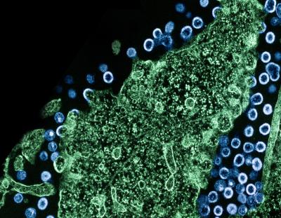
NIAID Scientists Detail First Structure of a Natural Mammalian Prion

NIAID Now |

NIAID Scientists Detail First Structure of a Natural Mammalian Prion

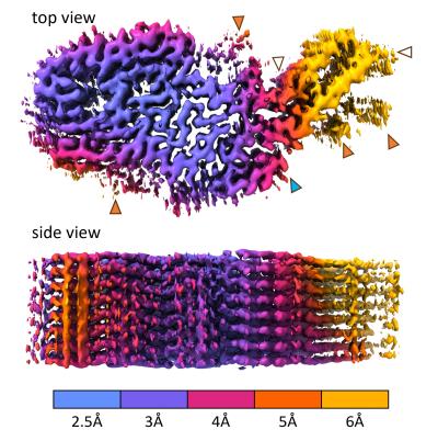
The near-atomic structure of a chronic wasting disease (CWD) prion should help scientists explain how CWD prions spread and become the most naturally infectious of the many mammalian protein aggregation diseases. NIAID scientists revealed the structure in a new study in Acta Neuropathologica. Such detailed knowledge could guide the rational design of vaccines and therapeutics, as well as identify mechanisms that protect humans from CWD pathogens in deer, elk, moose, and reindeer.

Many brain diseases of humans and other mammals involve specific proteins (e.g., prion protein or PrP) gathering into abnormal thread-like structures that grow by sticking to normal versions of the same protein. These threads can also fragment and spread throughout the nervous system and accumulate to deadly levels. For unknown reasons, CWD prions are more naturally contagious than most other protein aggregates and are spreading rampantly among cervid species in North America, Korea and northern Europe. Recalling the bovine spongiform encephalopathy (BSE) or “mad cow disease” epidemic of the mid-1980s and mid-1990s, there are concerns that CWD might similarly be transmissible to humans.

To date, no CWD transmission to humans has been substantiated, and the new CWD structure suggests preliminarily why we might be protected. The structure also reveals multiple differences between CWD and previously determined structures of highly infectious, but experimentally rodent-adapted, PrP-based prions. Differences are even more profound when compared to largely non-transmissible PrP filaments isolated from humans with Gerstmann-Sträussler-Scheinker syndrome, a genetic prion disorder.

PrP-based prion diseases are degenerative, untreatable, and fatal diseases of the central nervous system that occur in people and other mammals. These diseases primarily involve the brain, but also can affect the eyes and other organs. CWD-infected animals shed infectious prions in their feces, urine, and other fluids and body components while alive, and from their carcasses after dying. The prions can remain infectious in the environment for years. 

Scientists at NIAID’s Rocky Mountain Laboratories in Hamilton, Montana, determined the CWD structure from the brain tissue from a naturally infected white-tailed deer. They isolated the prions and froze them in glass-like ice. Then, using electron microscopy techniques, they developed a 3-D electron density map that indicated the detailed shapes of the protein molecules within the prion structure. This involved taking nearly 80,000 video clips of the sample, magnified 105,000 times the original size, at various orientations. They marked prion filaments in the video clips and collected more than 500,000 overlapping sub-images. They isolated about 7,300 of the highest quality sub-images and then used supercomputers to generate a 3-D density map and a molecular model to fit the map.

Vaccine development is among the many research areas where scientists could use high-resolution prion structures to advance their work. The study authors note that previous attempts to develop vaccines against CWD in cervids failed to be protective, and, at least in one case, had the opposite effect. They speculate that one explanation for adverse vaccine effects could be that antibody binding to the sides, rather than the ends of prion fibril surfaces, promotes fragmentation – creating infectious particles rather destroying them. Thus, a strategy to explore with vaccines and small-molecule inhibitors, they say, is to target the tips of prion structures where binding and conversion of prion protein molecules occurs.

The research team is planning to solve other naturally occurring prion structures, hoping to advance its understanding of the molecular basis of prion transmission and disease.

References:

P Alam, F Hoyt, E Artikis, et al. Cryo-EM structure of a natural prion: chronic wasting disease fibrils from deer. Acta Neuropathologica DOI: 10.1007/s00401-024-02813-y (2024).

A Kraus et al. High-resolution structure and strain comparison of infectious mammalian prionsMolecular Cell. DOI: 10.1016/j.molcel.2021.08.011. (2021).

Contact Information

Contact the NIAID Media Team.

301-402-1663
niaidnews@niaid.nih.gov

Search NIAID Blog

Save Your Future Self Time and Effort by Exploring ClinRegs Today

Funding News Editions:
See more articles in this edition

Governments set complex rules and regulations to ensure appropriate conduct of clinical trials. To aid researchers planning such studies around the globe, NIAID maintains the ClinRegs database of country-specific, clinical research regulatory information for 23 countries. For example, you could quickly check the clinical trial safety reporting requirements for Brazil; and if you have sites in other countries, you can compare safety reporting requirements side-by-side.

The best way for you to learn how to navigate the database is to watch Utilizing NIAID’s ClinRegs Website to Support International Clinical Research Regulatory Compliance, a webinar originally recorded on May 23, 2024. 

The ClinRegs team reviews country content yearly and makes interim updates to incorporate substantial, timely changes to country regulations or requirements. They also steadily upgrade the site’s features, recent examples of which include: 

  • A tabular dashboard on every country page that provides quick access to regulatory news, facts, current research and sites, recent page updates, and helpful links. 
  • An enhanced country page menu that includes pop-up descriptions of each topic and a topic filter for customized viewing. 
  • Comparison functionality that allows users to view up to four countries side-by-side. 
  • Filters to limit search results by country and topic, with an option to view results for multiple countries side-by-side. 

For screenshots highlighting these changes, refer to the February 2024 and May 2024 Functionality Upgrades pages. 

To receive the latest ClinRegs information, subscribe to country- or topic-specific email updates through GovDelivery. If you have any questions or comments, email the ClinRegs team at NIAIDClinRegsSupport@mail.nih.gov.

Contact Us

Email us at deaweb@niaid.nih.gov for help navigating NIAID’s grant and contract policies and procedures.

NIH Kicks Off Next Phase of U.S.-South Africa Program for Collaborative Biomedical Research

Funding News Editions:
See more articles in this edition

As part of the notice of funding opportunity (NOFO) U.S.-South Africa Program for Collaborative Biomedical Research—Phase 3 (HIV/AIDS) (R01, Clinical Trial Optional), NIAID, along with other NIH institutes and centers (ICs) and the South African Medical Research Council, will support the next phase of a collaborative program between U.S. and South African scientists carrying out HIV/AIDS research.

For Phase 3, NIAID is calling for applications to help foster, stimulate, and expand research on HIV/AIDS, HIV/AIDS comorbidities and co-infections, HIV/AIDS-associated implementation science, and HIV/AIDS-associated data science. The goal of this NOFO is to engage collaborative teams of U.S. and South African scientists working on these areas of research. 

Research Objectives 

As part of this NOFO, we encourage HIV-related research that’s in accordance with NIH’s overarching HIV research priorities as well as the NIH HIV/AIDS Research Priorities and Guidelines for Determining HIV/AIDS Funding.  

Our specific research areas of interest are as follows: 

  • Reduce incidence of HIV (prevention) 
  • Develop next-generation HIV therapies (treatment and care continuum) 
  • Research toward HIV cure 
  • Address HIV-associated comorbidities, co-infections, and complications 
  • Cancer 
  • Behavior, mental health, substance use, and HIV risk 
  • Implementation science/data science 

Refer to the NOFO for possible projects within each research area of interest.  

Note that we will consider applications proposing the following topics to be nonresponsive and not review them:  

  • Projects proposing Phase III or Phase IV Clinical Trials
  • Research using Select Agents
  • Applications without the required collaborative partnership (at least one South African program director/principal investigator (PD/PI) from an eligible institution from South Africa and at least one collaborator from a U.S. institution/organization). 

Who Can Apply 

Eligible National Research Foundation (NRF) South African Institutions, i.e., recognized South African public higher education or research institutions, can apply. (Refer to the NOFO for further details on eligible NRF South African institutions. Note that the South African Institution must submit the application, with the South African PI serving as the contact PI. The U.S. partner can serve as a PI on a multi-PI application or as a non-PI collaborator.)  

Application and Budget Information 

Applicant organizations may submit more than one application, provided that each application is scientifically distinct. Applicants will be required to include a data management and sharing plan that adheres to the NIH Grants Policy Statement.  

Issuing IC and partner components intend to fund 8 to 10 awards. Application budgets are not expected to exceed $400,000 in direct costs per year and must reflect the actual needs of the proposed project. While the maximum project period is 5 years, the scope of the proposed project should determine the project period.  

The application deadline is March 12, 2025. 

For more information, refer to Questions and Answers for U.S.-South Africa Program for Collaborative Biomedical Research—Phase 3 (HIV/AIDS). You can also contact Brian Remortel with NIAID-specific questions at remortelbg@niaid.nih.gov or 240-292-4816. Refer to the NOFO for contacts at other ICs.

Contact Us

Email us at deaweb@niaid.nih.gov for help navigating NIAID’s grant and contract policies and procedures.

NIAID Mouse Study Shows Existing Drug Could Limit COVID-19 Lung Damage

NIAID Now |

A drug that blocks danger signals that can lead to harmful inflammation could help reduce COVID-19 lung damage, a new study from NIAID scientists and colleagues has found. Scientists from NIAID’s Rocky Mountain Laboratories in Hamilton, Montana, and the University of Utah completed the project, published online in JCI Insights.

Though they completed the study in laboratory mice modified to model COVID-19 in people, the scientists think their findings are important enough to pursue further studies of the drug, FPS-ZM1, to determine dosing and timing strategies for possible human clinical trials. FPS-ZM1 is an immune modulatory therapeutic – the drug is designed to prevent a specific immune system response from occurring. The investigational therapy has been evaluated in preclinical studies to treat conditions such as diabetes, lung injury and stroke. In their study, the scientists used FPS-ZM1 to block the “receptor for advanced glycation end products” (RAGE), which senses danger signals and can generate inflammation and coagulation known to damage the lungs of COVID-19 patients. 

Therapeutic treatment with FPS-ZM1 during the study improved survival in mice infected with SARS-CoV-2, the virus that causes COVID-19. Further, FPS-ZM1 specifically reduced damage to the lung vasculature, an important system for circulating blood through the lungs that becomes damaged during SARS-CoV-2 infection. FPS-ZM1 also has shown in other rodent studies that it can protect against injury in disease models of brain injury, sepsis, asthma, diabetes, acute lung injury and ischemic/reperfusion (organ damage due to blood flow).

The study also identified two distinct phases of COVID-19 disease development in the mice. The scientists want to further explore those phases as potential guides for treatment strategies. For example, FPS-ZM1 limited specific types of inflammation and tissue damage, so it would likely be most effective if administered during the intermediate to later stages of SARS-CoV-2 infection, whereas antiviral treatment may be most effective when given early following infection.

Reference: F Jessop, et al. Impairing RAGE signaling promotes survival and limits disease pathogenesis following SARS-CoV-2 infection. JCI Insights DOI: https://doi.org/10.1172/jci.insight.155896. (2022).
 

Contact Information

Contact the NIAID Media Team.

301-402-1663
niaidnews@niaid.nih.gov

Search NIAID Blog

COVID-19 Respiratory Treatment Effective in Encephalitis Study

NIAID Now |

COVID-19 Respiratory Treatment Effective in Encephalitis Study

Molnupiravir Reduced Viral Brain Disease in Mice

NIAID research into finding broad uses for existing drug treatments has a potential new success story: Molnupiravir, a relatively new antiviral developed to treat respiratory diseases – such as COVID-19 – reduced brain swelling in study mice infected with a pathogen dangerous to children, La Crosse virus (LACV).

The new study, from NIAID scientists and colleagues at the University of North Carolina and Emory University, is published in PLOS Pathogens. LACV, which is spread by mosquitos, can cause brain inflammation in children. LACV was first isolated in the early 1960s near La Crosse, Wisconsin. Since then, LACV encephalitis cases have been found in more than 20 states, mostly in the basins of the Mississippi and Ohio rivers and throughout the Appalachian Mountains.

Most LACV infections in people are mild, but the virus sometimes – particularly in children – enters the brain, infects neurons and causes disease that can result in learning and memory difficulties, paralysis, seizures and death. Between 30 and 90 cases of severe LACV – those that affect the central nervous system (CNS) – are reported each year, though the Centers for Disease Control and Prevention believes many mild cases occur but are not diagnosed.

The study used a new strategy to test three antiviral drugs – N4-Hydroxycytidine (NHC, the active metabolite of the prodrug molnupiravir), ribavirin and favipiravir– for treatment against LACV infection. The scientists chose LACV because it broadly represents several RNA viruses that cause disease in the CNS, including Jamestown Canyon and Cache Valley viruses – which also were part of the study – and rabies, polio, West Nile, Nipah and several other viruses not part of the study.

The three antiviral drugs were tested in a cell culture system to examine an antiviral strategy called lethal mutagenesis. This approach increases the number of errors in the viral genome that RNA viruses make when they replicate, weakening the resulting viruses. By incorporating the drug, more errors are induced in the viral genome and more weakened viruses emerge, providing the host an opportunity to recover.

Ribavirin and favipiravir used in cell treatment studies did not produce potent enough results to justify testing in mice. The NHC prodrug molnupiravir, however, was used in two different mouse study models. Oral treatment with molnupiravir reduced brain disease in mice by 32% when LACV infection was started by an injection in the abdomen, and by 23% when the infection was started in the nose, offering easy access to the brain.

Also noteworthy: The researchers tested NHC against LACV and found it effective in the cell and mouse models, as well as in cell models using Jamestown Canyon and Cache Valley viruses. This showed that the drug treatment strategy could be successful against viruses related to LACV and supports the idea that this strategy could be used against a broader group of encephalitic RNA viruses.

The researchers say more study is needed to see how these drugs counter RNA viruses, particularly to determine whether injecting the drug directly into cerebrospinal fluid would provide better results and possibly reduce adverse side effects.

Reference:

D Ojha et alN4-Hydroxycytidine/Molnupiravir Inhibits RNA Virus-Induced Encephalitis by Producing Less Fit Mutated VirusesPLOS Pathogens DOI: 10.1371/journal.ppat.1012574 (2024).

Contact Information

Contact the NIAID Media Team.

301-402-1663
niaidnews@niaid.nih.gov

Search NIAID Blog

Childhood Pneumonia Study Shows Short-Course Antibiotics Superior to Standard of Care

The Eyes Have it: A Functional Role for Prion Protein

NIAID Now |

In the early 1980s, scientists identified clumps of abnormal, misfolded prion protein in mammals as the cause of brain-wasting diseases, now called prion diseases. Since that time, they have struggled to answer: What does a normal prion protein do?

The answer, they believe, could help lead them to develop treatments and disease-prevention measures against human prion diseases, such as Creutzfeldt-Jakob disease, fatal familial insomnia and kuru, as well as animal prion diseases, such as scrapie in sheep and chronic wasting disease in cervids.

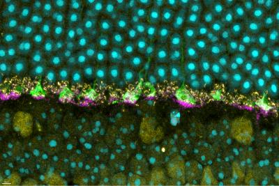
Now, a new study published in iScience from NIAID scientists at Rocky Mountain Laboratories in Hamilton, Montana, and colleagues provides details of how prion protein functions in the retina of mouse eyes, helping them respond to light.

The scientists used mice specially bred without prion protein to compare to wild mice with natural prion protein. Prior studies have suggested that prion protein may have a role in how nerves transmit signals to other nerves at specialized junctions, known as neural synapses. So, knowing that prion protein exists naturally in the eye, the researchers examined mouse retina for a specific neural synapse role.

A key tool the researchers used involved measuring the electroretinographic (ERG) responses – the amount of time it took for the retina in mice to respond to a flash of light. Remember as a kid in school learning about rods and cones in the eye and how they convert light signals to help the brain understand vision? The same is true in mice.

Compared to the wild mice with prion protein, the scientists observed deficiencies in ERG responses for mice without prion protein. The deficiencies affected the normal function of the rods and cones. And – using the ERG data and neural synapse information – they found that the deficiencies originated in the portion of the retina where natural prion protein was most highly concentrated.

Though additional study is needed, the researchers believe the prion protein may act like scaffolding to help cells and elements of the eye, such as rods and cones, to stabilize neural synapses. And they believe prion protein must be present for rods and cones to function normally.

The research team hopes these findings help colleagues who study prion diseases better understand what might occur in humans when natural forms of prion protein are therapeutically removed. New treatment strategies for prion diseases focus on using drugs that remove natural prion protein to eliminate the potential for misfolding and clumping. But researchers do not know whether that could result in unwanted outcomes, such as possibly affecting vision. These findings also could extend to other protein-related neurodegenerative diseases, such as Alzheimer’s (amyloid beta protein) and Parkinson’s diseases (alpha synuclein protein).

Scientists from Duke University and the McLaughlin Research Institute in Great Falls, Montana, collaborated on the study.

Reference: J Striebel, et al. The prion protein is required for normal responses to light stimuli by photoreceptors and bipolar cells. iScience DOI: 10.1016/j.isci.2024.110954 (2024).

Contact Information

Contact the NIAID Media Team.

301-402-1663
niaidnews@niaid.nih.gov

Search NIAID Blog

NIH Releases Mpox Research Agenda

The NIAID mpox research agenda focuses on four key objectives: increasing knowledge about the biology of all clades—also known as strains—of the virus that causes mpox, including how the virus is transmitted and how people’s immune systems respond to it; evaluating dosing regimens of current vaccines to stretch the vaccine supply and developing novel vaccine concepts; advancing existing and novel treatments, including antivirals and monoclonal antibodies; and supporting strategies for detecting the virus to facilitate clinical care and epidemiological surveillance.

Contact

Submit a Media Request

Contact the NIAID News & Science Writing Branch.

301-402-1663
niaidnews@niaid.nih.gov
All Media Contacts

Study Links Certain Vaginal Bacteria and Inflammatory Marker to Increased Odds of Acquiring HIV Among Cisgender Women

NIAID Now |

Fourteen vaginal bacterial species and the presence of a protein that promotes inflammation were associated with increased odds of HIV acquisition in a study of more than 500 cisgender women in African countries with high HIV incidence. The study was the largest to date to prospectively analyze the relationship between both the vaginal microbiome and vaginal tissue inflammation and the likelihood of acquiring HIV among cisgender women in this population. The NIAID-sponsored research was published in The Journal of Infectious Diseases.

Research is limited regarding the potential impacts of vaginal bacteria and inflammatory markers on HIV acquisition. Only one previous study has characterized both factors in women before they had HIV to investigate their odds of acquiring the virus, but the number of HIV acquisition events in that study was low, potentially limiting their ability to detect associations.

To increase understanding of these issues, researchers analyzed vaginal swab samples from 586 cisgender women participating a large biomedical HIV prevention clinical trial in South Africa, Uganda and Zimbabwe, and compared the bacterial and inflammatory profiles of samples from 150 participants who acquired HIV during the study with the samples of 436 participants who did not. The team identified 14 bacterial species associated with HIV acquisition and noted that participants whose samples contained most or all of those bacteria had the highest odds of acquiring HIV, while the presence of none or few of the identified bacteria was associated with the lowest odds of HIV acquisition. They similarly identified six inflammatory cytokines and chemokines—proteins that communicate with other cells to prompt the body to fight infections through inflammatory processes—associated with HIV acquisition, and identified the highest odds of HIV acquisition in participants whose samples contained all six of those proteins. Furthermore, they identified a single chemokine called interferon gamma-induced protein 10 associated with the highest odds of HIV acquisition out of the six.

These results suggest that strategies to reduce concentrations of the 14 identified bacterial species and inflammatory proteins could help prevent HIV acquisition, according to the authors. They also recommended that additional studies be conducted to understand the mechanisms by which these factors contribute to biological susceptibility to HIV.

Reference: Srinivasan, S et al. Vaginal Bacteria and Proinflammatory Host Immune Mediators as Biomarkers of HIV Acquisition 3 Risk among African Women. Journal of Infectious Diseases. DOI 10.1093/infdis/jiae406 (2024).

Contact Information

Contact the NIAID Media Team.

301-402-1663
niaidnews@niaid.nih.gov

Search NIAID Blog

USask’s VIDO Awarded NIH Contract to Develop Critical Models for Global Research

Publish or Event Date
Research Institution
University of Saskatchewan
Short Title
USask’s VIDO Awarded NIH Contract to Develop Critical Models for Global Research
Content Coordinator
Content Manager