The immunology team interrogates immune responses against pathogens requiring maximum containment by use of a variety of capabilities. These include but are not limited to cytometry (e.g., immunophenotyping), Luminex multiplex protein/cytokine arrays, cartridge-based cell- sorting (MACSQuant Tyto), ELISAs, serology, in vitro functional assays, intracellular cytokine staining (ICS) enzyme-linked immune absorbent spot (ELISPOT; e.g., T cell and B cell).
Main Areas of Focus
- Immunophenotyping and immune kinetics
- Pathogen-specific serology
- Multiplex cytokine assays and analysis
- In vitro functional assays
Capabilities and Specialized Equipment
- Custom pathogen-specific assay development
- Pathogen-specific prescreening
- Data interpretation and analysis
- BD LSRFortessa flow cytometers with four lasers (405 nm, 488 nm, 561 nm, 640 nm) (one at BSL-4, one at BSL-2) and up to 16 fluorescent parameters
- Luminex Flexmap 3D Instrument System (one at BSL-4)
- Cytek Aurora flow cytometer with five lasers (355 nm, 405 nm, 488 nm, 561 nm, 640 nm) (two at BSL-4, two at BSL-2) and up to 63 simultaneous fluorescent parameters
- ONI Nanoimager S super-resolution microscope with four lasers (405 nm, 488 nm, 561 nm, 640 nm) for PALM, dSTORM, smFRET, and 3D single-particle tracking (one planned to go into BSL-4)
- Miltenyi Biotec MACSQuant Tyto cartridge-based cell sorter with three lasers (405 nm, 488 nm, 638 nm) (one planned to go into BSL-4) (Low pressure: 3-psi cartridge, 18-psi high-speed cartridge) with up to eight simultaneous fluorescent parameters
- Miltenyi Biotec gentleMACS Octo Dissociators with Heaters (two at BSL-4)
- Tecan Infinite M200 PRO multimode plate reader (absorbance, fluorescence, luminescence)
- AquaMax 2000 plate washer with magnetic and cell-washing capabilities
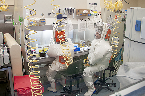
Erika and Tony work in the biosafety cabinet within the BSL-4 laboratory to isolate and stain cells attained from tissues collected during necropsy; subsequently, the cells will be analyzed via flow cytometry.
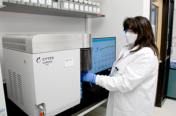
Rebecca processes a stained and fixed whole-blood sample on the Cytek Aurora Flow Cytometer in the BSL-2 laboratory.

Tony prepares a chamber slide in the BSL-2 laboratory. These slides are used for imaging with the ONI Nanoimager S super-resolution microscope.
Location
Integrated Research Facility at Fort Detrick (IRF-Frederick)
Contact Information
Jens H. Kuhn, M.D., Ph.D., Ph.D., M.S.
Principal Scientist and Director of Virology (Contractor)
Gabriella Worwa, D.V.M.
Associate Supervisor and Study Director (Contractor)
Bapi Pahar D.V.M., M.V.Sc., Ph.D., M.B.A.
Immunologist (Contractor)
IRF-Frederick
Standards
All procedures are well-documented and adhere to standard operating procedures (SOPs), methods, or study-approved plans and agreements.
Collaboration Opportunities
- Studies relevant to human disease
- Use of surrogate systems to test clinical hypotheses
- Use of biological systems to answer questions regarding disease pathogenesis and strategies for intervention including antimicrobials, vaccines, and other countermeasures
- Developing and incorporating cutting-edge technologies to understand infectious diseases
Read more about how to work with the IRF-Frederick.