The pathology and histology teams work together to conduct morphologic assessments and molecular assays using in-life and postmortem samples attained during research studies that develop and use established animal models of high-consequence emerging human viruses. The clinical pathologic and histopathologic data are interpreted by the pathologists using historical controls, or within-study controls, to generate reports that address specific pathology-associated aims of each study. The findings facilitate the development of future studies and contribute to publications in high-quality peer-reviewed journals.
Main Areas of Focus
- Participate in study planning to ensure successful execution of pathology-related specific aims (i.e., appropriate animal model selection, assay selection, and sample allocation)
- Conduct postmortem examinations in laboratory animal models under biosafety level 4 (BSL-4) conditions
- Review protocol development of molecular assays to detect emerging viral pathogens and relevant biomarkers in tissue in collaboration with our histotechnologists
- Interpret clinical pathology data
- Interpret cytology findings in blood smears and cytospin samples (i.e., bronchoalveolar lavage fluid, cerebrospinal fluid, and urine)
- Interpret histopathologic lesions in tissue sections
- Interpret immunohistochemical (IHC) findings in tissue sections
- Interpret in situ hybridization (ISH) findings in tissue sections
- Conduct histopathology scoring based on lesion type or staining characteristics in tissues to generate semiquantitative histopathology data for (statistical) significance testing
- Conduct and interpret electron microscopic tissue examinations
- Produce publication-quality pathology reports
- Contribution to the writing and publication of manuscripts and present pathology data
Capabilities and Specialized Equipment
- Full-service gross dissection, processing, embedding, microtomy, staining, and coverslip application
- Mopec MB-600 adjustable elevating grossing station with backdraft ventilation
- Tissue-Tek VIP-6 automated thermoregulated vacuum infiltration processor
- Tissue-Tek TEC-6 ergonomic embedding console and cryo module workstation
- Leica Biosystems HistoCore-MultiCut standard semiautomated rotary microtome
- Leica Biosystems HI1210 lighted thermoregulated water flotation bath
- Tissue-Tek/Sakura-Finetek Prisma Plus and Glas g2 automated slide stainer and coverslip applicator
- Leica Biosystems IP-C/IP-S chemical-resistant inkjet printers for cassettes/slides
- Immunohistochemistry (IHC)
- ThermoScientific-Shandon Sequenza staining system, chromogenic/fluorescent
- In situ hybridization (ISH)
- Advanced Cell Diagnostics-BioTechne RNAscope HybEZ-II staining system
- Digital pathology whole-slide image scanning and biomarker quantitation
- Leica Aperio AT2 high-throughput digital slide scanner
- Publication-quality image capture of brightfield photomicrographs
- Olympus BX Series multimodule microscope and integrated DP74 camera
- Publication-quality image capture of fluorescent photomicrographs
- Leica SP5 TCS-2 confocal microscope and integrated DMI6000 camera with five internal fluorescent detectors, two HyD high-sensitivity detectors, and transmitted light detector with laser lines 405, 458, 476, 488, 514, 561, 594, and 633
- Leica SP5 TCS-2 confocal microscope and integrated DMI6000 camera with five internal fluorescent detectors, two HyD high-sensitivity detectors, and transmitted light detector with laser lines 405, 458, 476, 488, 514, 561, 594, and 633
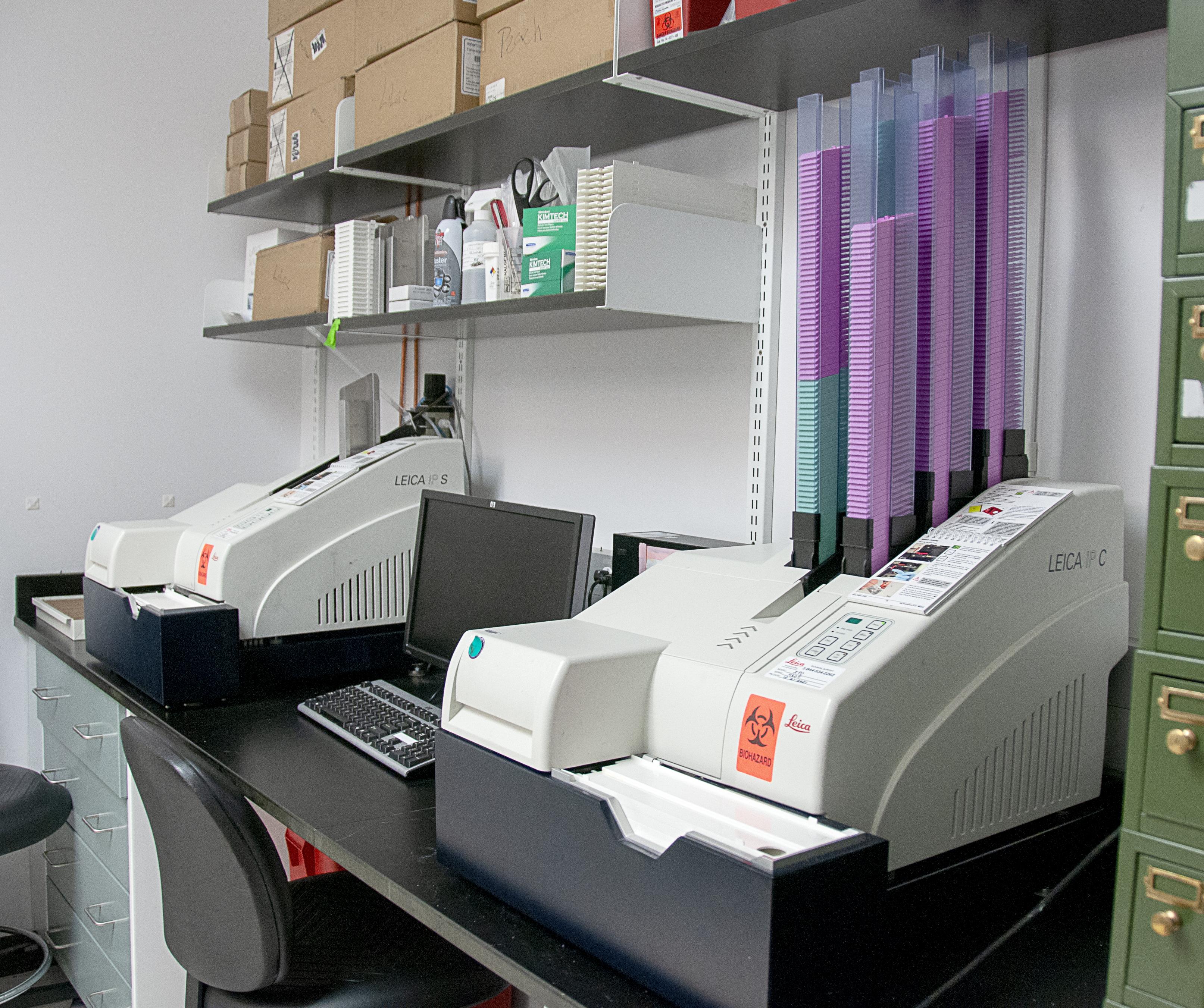
Leica Biosystems IP-C/IP-S chemical-resistant inkjet printers for cassettes and slides.
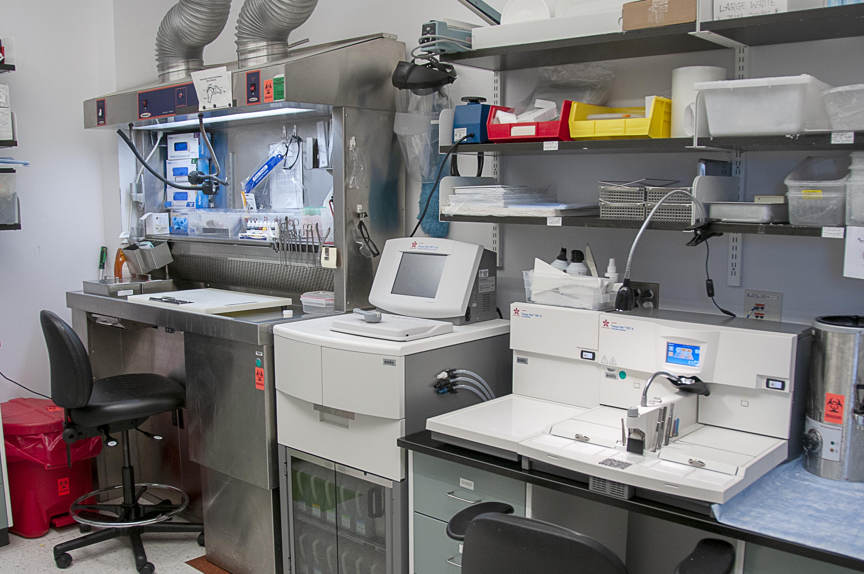
Mopec MB-600 adjustable elevating grossing station with backdraft ventilation, Tissue-Tek VIP-6 automated thermoregulated vacuum infiltration processor unit, and Tissue-Tek TEC-6 ergonomic embedding console and cryo module workstation.
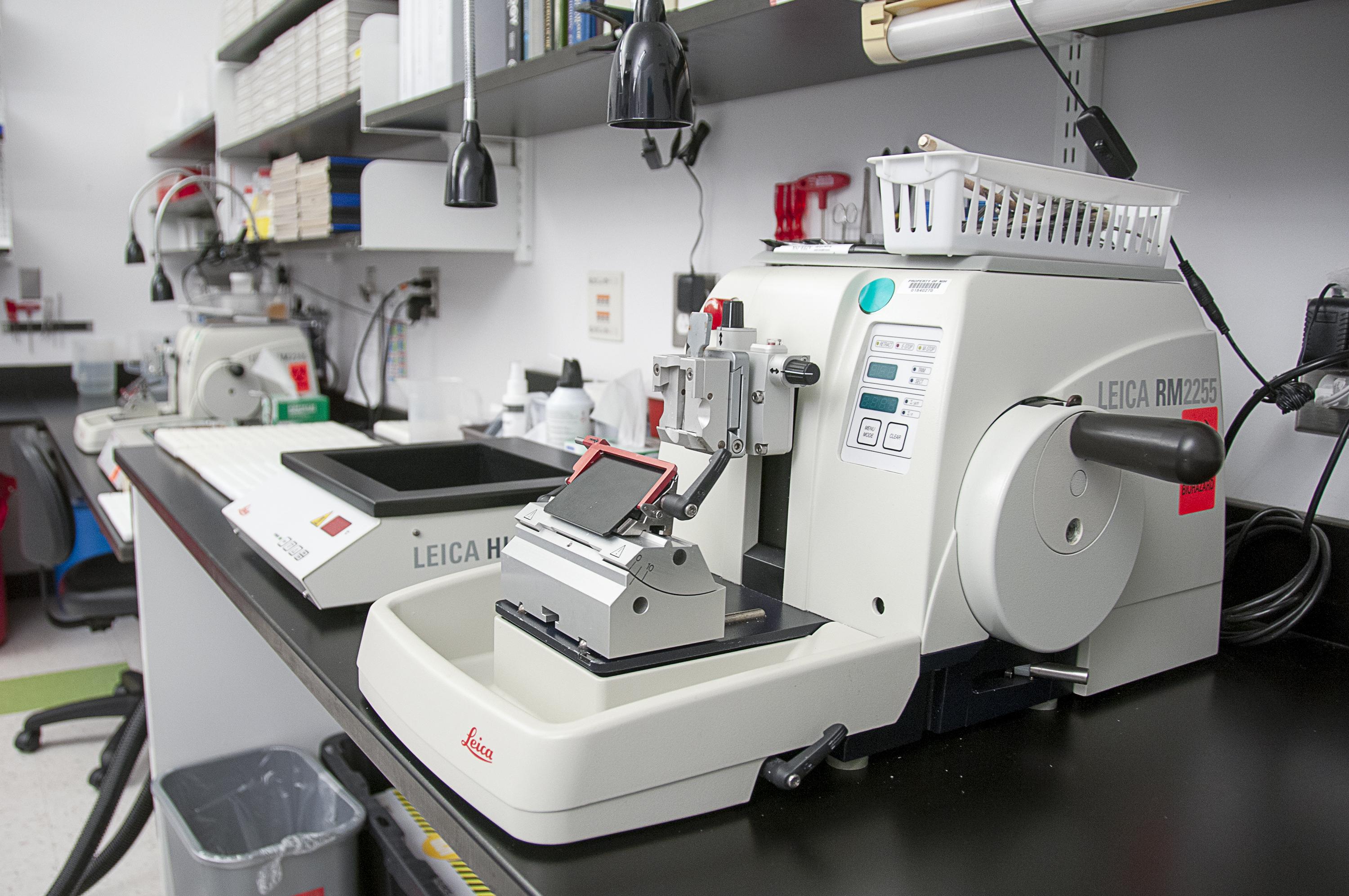
Leica Biosystems HistoCore-MultiCut standard semiautomated rotary microtome and HI1210 lighted thermoregulated water flotation bath instrument.
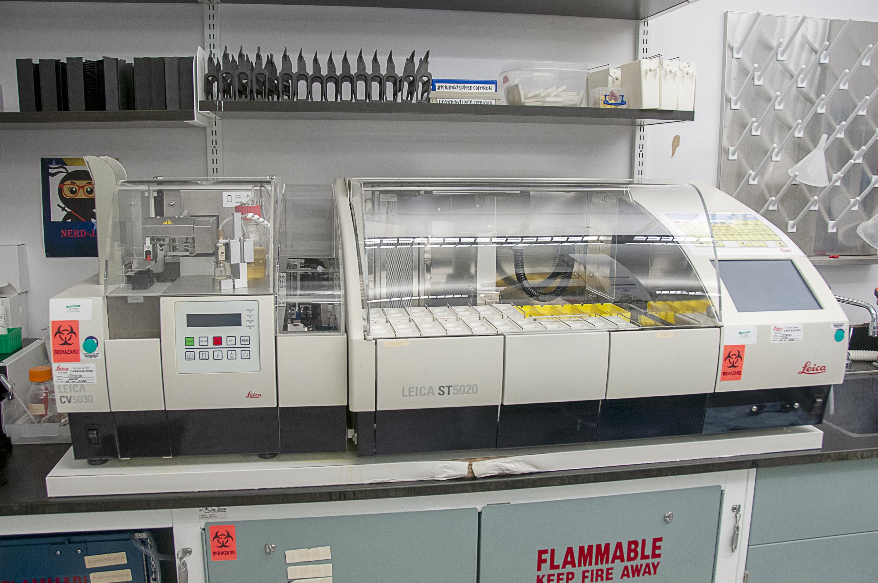
Leica Biosystems ST5020/CV5030 integrated autostainer and coverslip applicator.
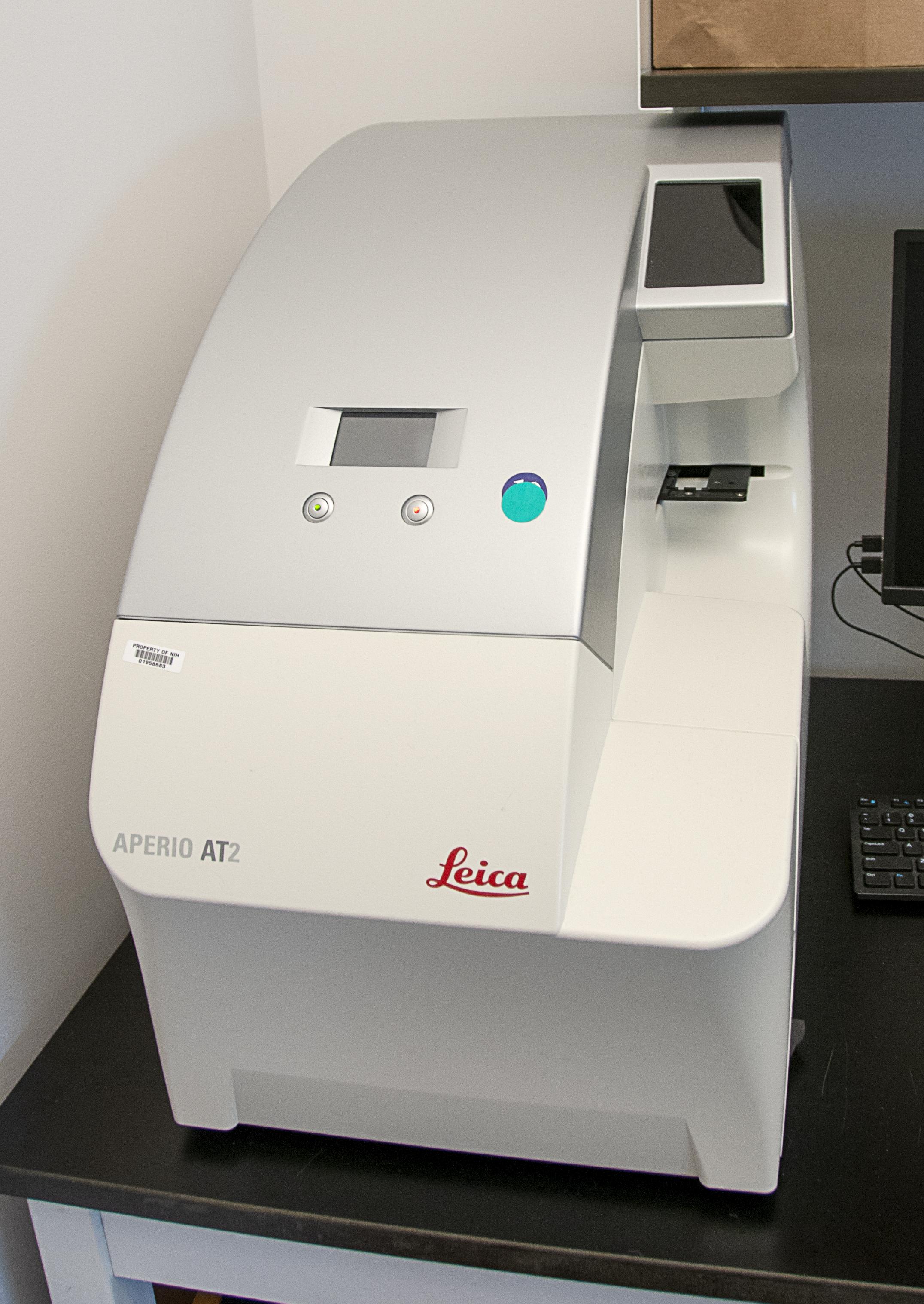
Leica Aperio AT2 high-throughput digital slide scanner.

Leica SP5 TCS-2 confocal microscope and integrated DMI6000 camera system with five internal fluorescent detectors, two HyD high sensitivity detectors, and transmitted light detector with laser lines 405, 458, 476, 488, 514, 561, 594, and 633.
Location
Integrated Research Facility at Fort Detrick (IRF-Frederick)
Contact Information
Venkatesh Mani, Ph.D.
Director of Imaging and Maximum Containment (Contractor)
Louis Huzella, D.V.M., Diplomate, ACVP
Pathology Lead (Contractor)
IRF-Frederick
Standards
All procedures are well-documented and adhere to standard operating procedures (SOPs), methods, or study-approved plans and agreements.
Collaboration Opportunities
- Studies relevant to human disease
- Use of surrogate systems to test clinical hypotheses
- Use of biological systems to answer questions regarding disease pathogenesis and strategies for intervention including antimicrobials, vaccines, and other countermeasures
- Developing and incorporating cutting-edge technologies to understand infectious diseases
Read more about how to work with the IRF-Frederick.