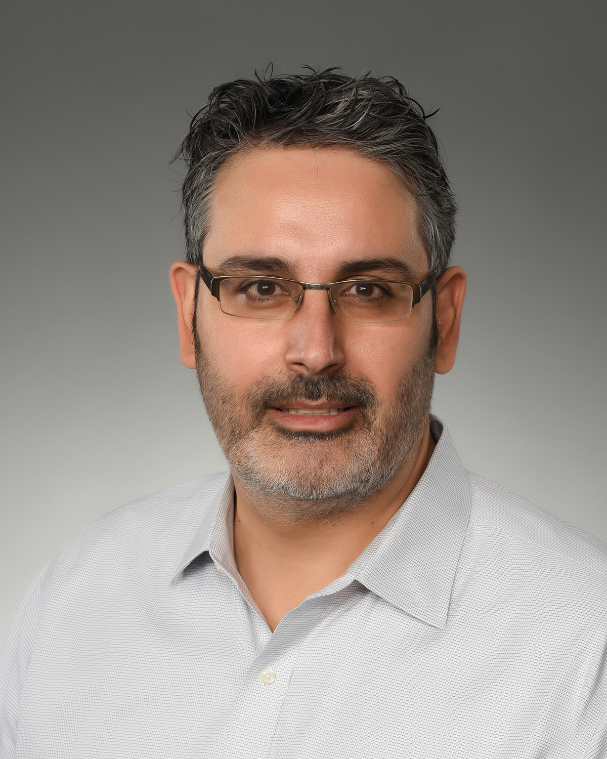
Neeltje van Doremalen, Ph.D.

Section or Unit Name
Mucosal Immunology and Virology Unit (MIVU)
Exclude from directory
Off
Section/Unit: Year Established
Section/Unit: Location
This Researcher/Clinician’s Person Page
Parent Lab/Program
Program Description

Our lab is dedicated to understanding the unique role of the mucosal immune system in protecting the respiratory tract against viral infections. Unlike the systemic immune system, the mucosal immune system acts as the first line of defense at critical surfaces such as the respiratory tract, gut, and reproductive organs. Key players in this defense include tissue-resident memory T cells and secretory IgA, which operate independently of systemic responses. We aim to unravel how mucosal immunity is induced and how it provides protection against respiratory viruses, particularly in the context of infections in the upper and lower respiratory tracts.

We investigate the immune responses elicited by respiratory viruses such as influenza A viruses and coronaviruses, focusing on both mucosal and systemic adaptive immunity. Using rodent models, we study the humoral and cellular responses elicited by different infection routes and analyze the role of innate immunity in shaping adaptive responses. By leveraging techniques like high dimensional flow cytometry, systems serology, deep mutational scanning, single-cell transcriptomics, and multiplex imaging, we gain spatial and temporal insights into immune responses across critical tissues, including the nasal-associated lymphoid tissue, nasal turbinates, lungs, and lymph nodes. These studies enable us to map mucosal immunity comprehensively and identify the breadth and depth required for protection upon rechallenge.

We additionally aim to identify correlates of protection and optimize vaccine strategies to induce robust mucosal immunity. We evaluate diverse vaccine platforms—including mRNA, vectored, and subunit vaccines—administered via various routes, such as intranasal, intramuscular, and inhalation. By comparing vaccine technologies and regimens, we aim to establish principles for designing universal vaccines capable of inducing broad, durable mucosal immune responses. Ultimately, our goal is to provide foundational insights that improve vaccine design and our understanding of protective mechanisms against respiratory viruses.

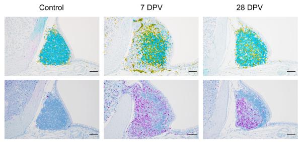
Immunohistochemistry staining of nasal-associated lymphoid tissues (NALT). The upper panels show CD3 (yellow, marking T cells) and PAX5 (teal, marking B cells). The lower panels depict Ki67 (purple, marking proliferating cells).

Immunohistochemistry staining of nasal-associated lymphoid tissues (NALT). The upper panels show CD3 (yellow, marking T cells) and PAX5 (teal, marking B cells). The lower panels depict Ki67 (purple, marking proliferating cells). Samples were collected at various time points following intranasal vaccination of mice with a replication-incompetent adenovirus vaccine. Acknowledgements: Reshma K. Mukesh, Carl Shaia, Jessy Prado-Smith.

Credit: NIAID
Immunohistochemistry staining of nasal turbinates and NALT tissues with CD3 (brown), highlighting the migration of T cells into these regions.

Immunohistochemistry staining of nasal turbinates and NALT tissues with CD3 (brown), highlighting the migration of T cells into these regions. Samples were collected at various time points after intranasal vaccination of mice with a replication-incompetent adenovirus vaccine. Acknowledgements: Reshma K. Mukesh, Carl Shaia, Jessy Prado-Smith.

Credit: NIAID
Selected Publications

Cohen AA, van Doremalen N, Greaney AJ, Andersen H, Sharma A, Starr TN, Keeffe JR, Fan C, Schulz JE, Gnanapragasam PNP, Kakutani LM, West AP Jr, Saturday G, Lee YE, Gao H, Jette CA, Lewis MG, Tan TK, Townsend AR, Bloom JD, Munster VJ, Bjorkman PJ. Mosaic RBD nanoparticles protect against challenge by diverse sarbecoviruses in animal models. Science. 2022 Aug 5;377(6606):eabq0839.

van Doremalen N, Purushotham JN, Schulz JE, Holbrook MG, Bushmaker T, Carmody A, Port JR, Yinda CK, Okumura A, Saturday G, Amanat F, Krammer F, Hanley PW, Smith BJ, Lovaglio J, Anzick SL, Barbian K, Martens C, Gilbert SC, Lambe T, Munster VJ. Intranasal ChAdOx1 nCoV-19/AZD1222 vaccination reduces viral shedding after SARS-CoV-2 D614G challenge in preclinical models. Sci Transl Med. 2021 Aug 18;13(607):eabh0755.

Holbrook MG, Anthony SJ, Navarrete-Macias I, Bestebroer T, Munster VJ, van Doremalen N. Updated and Validated Pan-Coronavirus PCR Assay to Detect All Coronavirus Genera. Viruses. 2021 Apr 1;13(4):599.

van Doremalen N, Lambe T, Spencer A, Belij-Rammerstorfer S, Purushotham JN, Port JR, Avanzato VA, Bushmaker T, Flaxman A, Ulaszewska M, Feldmann F, Allen ER, Sharpe H, Schulz J, Holbrook M, Okumura A, Meade-White K, Pérez-Pérez L, Edwards NJ, Wright D, Bissett C, Gilbride C, Williamson BN, Rosenke R, Long D, Ishwarbhai A, Kailath R, Rose L, Morris S, Powers C, Lovaglio J, Hanley PW, Scott D, Saturday G, de Wit E, Gilbert SC, Munster VJ. ChAdOx1 nCoV-19 vaccine prevents SARS-CoV-2 pneumonia in rhesus macaques. Nature. 2020 Oct;586(7830):578-582.

Folegatti PM, Ewer KJ, Aley PK, Angus B, Becker S, Belij-Rammerstorfer S, Bellamy D, Bibi S, Bittaye M, Clutterbuck EA, Dold C, Faust SN, Finn A, Flaxman AL, Hallis B, Heath P, Jenkin D, Lazarus R, Makinson R, Minassian AM, Pollock KM, Ramasamy M, Robinson H, Snape M, Tarrant R, Voysey M, Green C, Douglas AD, Hill AVS, Lambe T, Gilbert SC, Pollard AJ; Oxford COVID Vaccine Trial Group. Safety and immunogenicity of the ChAdOx1 nCoV-19 vaccine against SARS-CoV-2: a preliminary report of a phase 1/2, single-blind, randomised controlled trial. Lancet. 2020 Aug 15;396(10249):467-478.

van Doremalen N, Bushmaker T, Morris DH, Holbrook MG, Gamble A, Williamson BN, Tamin A, Harcourt JL, Thornburg NJ, Gerber SI, Lloyd-Smith JO, de Wit E, Munster VJ. Aerosol and Surface Stability of SARS-CoV-2 as Compared with SARS-CoV-1. N Engl J Med. 2020 Apr 16;382(16):1564-1567.

Visit PubMed for a complete publication listing.

Major Areas of Research
  • Understanding mucosal immunity induced by respiratory virus infections and mucosal vaccination
  • Identifying correlates of protection against respiratory virus infections
  • Utilize this knowledge to design improved vaccines

Candida auris—a mysterious and tenacious enemy

NIAID Now |

Certain species of fungi are responsible for many common infections including yeast infections, ringworm, thrush, and athlete’s foot. While these diseases may not lead to serious outcomes for most healthy individuals, fungal infections can be deadly, especially for patients with weakened immune systems. One fungal pathogen, Candida auris, is an emerging healthcare-associated infection of growing public health concern. The first case of Candida auris was reported in 2009 in Japan, where it was isolated from a patient’s ear (Auris is Latin for “ear”). Outbreaks have since emerged rapidly around the globe. In the United States, C. auris infections have been increasing over the past few years, with more than 1460 cases reported in 2021. C. auris is typically found in hospitals and other healthcare settings and can cause serious bloodstream and wound infections.

Like other Candida species, C. auris is a type of yeast. However, unlike its yeasty cousins, this pathogen can colonize patients’ skin and persist for long periods of time on environmental surfaces. Another challenge is that C. auris is often resistant to one or more of the major classes of drugs that are typically used to treat fungal infections. While most C. auris infections can be treated with a class of antifungals called echinocandins, resistance to these drugs has also been reported, making some infections difficult to treat. C. auris is the only fungal pathogen identified as an ‘urgent’ threat in CDC’s Antibiotic Resistance Threat Report.

Back to basics

NIAID supports several researchers who are asking fundamental questions about the biology of C. auris. Today’s NIAID Now post features insights from NIAID-funded researchers, Jeniel Nett, M.D., Ph.D., associate professor of medicine and medical microbiology and immunology at University of Wisconsin-Madison, and Christina Cuomo, Ph.D., associate director of the Genomic Center of Infectious Diseases at the Broad Institute of Massachusetts Institute of Technology and Harvard University.

How is C. auris able to colonize the skin and persist in the environment?

C. auris can live and grow on the skin or in the body without causing illness. However, people who are colonized with C. auris may spread the pathogen to others and are at risk of getting sick later on if they develop infections. One important question to understanding C. auris outbreaks is: how is the fungus able to colonize skin so effectively and to persist in the environment? Dr. Nett’s research group is tackling this question by studying C. auris growth in the lab using two different systems. The first is designed to mimic human sweat and skin. Nett noted, “we think that this represents skin to some degree but also when surfaces get contaminated with skin and sweat components.” The other system is pig skin. “Pigs have similar skin to humans in terms of skin thickness and some of the cell types,” Nett explained. Using these systems, Nett and colleagues have shown that C. auris is able to readily grow on skin. “It really seems to mirror what we’re seeing patients,” said Nett. They’ve found that when the fungus is grown in the synthetic sweat medium, it forms multi-layered plaques, or biofilms, both on the pig skin as well as on hard surfaces. Compared to other Candida species, the biofilms are thicker and contain more viable organisms. C. auris biofilms can also persist on surfaces without drying out for up to two weeks in the lab.

A professional headshot of Dr. Nett standing in front of a window. She has long blonde hair, and is wearing a lab coat.

Jeniel Nett, M.D., Ph.D., associate professor of medicine and medical microbiology and immunology at University of Wisconsin-Madison

Credit: Dr. Jeniel Nett

Nett’s research demonstrates the ability of the fungus to colonize skin and form persistent biofilms on environmental surfaces, which has implications for transmission in healthcare settings. “This really becomes important with reusable medical equipment that goes room to room,” Nett emphasized. The systems Nett’s group has developed to study C. auris in the lab can also inform potential strategies to remove C. auris from the skin of patients. Nett’s research has shown that while antiseptics are somewhat effective, they are not as active against C. auris when the fungus is growing in the skin environment compared to when it is growing without the skin present. In a published manuscript, Nett and colleagues demonstrated that the commonly used topical antiseptic chlorhexidine does not fully remove C. auris from the skin of patients. They also showed that by adding isopropanol, as well as some essential oils, including tea tree and lemongrass, to chlorhexidine, they were able to improve the activity of the antiseptic. Her group is still investigating what specific components of skin and sweat are triggering biofilm growth in C. auris. Understanding this could lead to better, more specific strategies to disrupt skin colonization.

How did C. auris outbreaks emerge around the world, and how has the fungus become multidrug-resistant?

Soon after it was first identified, outbreaks of C. auris arose in four distinct locations—South Asia, East Asia, Africa, and South America—nearly simultaneously. Dr. Cuomo and colleagues are using a genomics approach to better understand this phenomenon. 
“One fundamental question genomics can answer is, what has been the history of the pathogen over time?” Cuomo explained. “We can take isolates from different patients, and by comparing them we can infer back in time to where they have a common connection.”

A professional headshot of Dr. Cuomo, standing in a bright atrium. She has short brown hair and is wearing glasses.

Christina Cuomo, Ph.D., associate director of the Genomic Center of Infectious Diseases at the Broad Institute of Massachusetts Institute of Technology and Harvard University

Credit: Dr. Christina Cuomo

Together with colleagues at the Centers for Disease Control and Prevention, Cuomo’s group helped confirm that the different outbreaks were caused by distinct genetic groups, or ‘clades.’ As cases have continued to spread around the globe, researchers have been able to trace new C. auris isolates back to these four major clades, allowing them to understand how the different outbreaks are connected.

Expanding on this initial work, Cuomo’s group is looking more closely at the different C. auris clades and identifying key genetic differences both within and between these groups as well as among C. auris and other related Candida species. From this analysis, they have generated hypotheses about which genes in the fungus are important for contributing to disease in humans. Such studies provide important insight into the biology of C. auris and can help identify potential targets for new drugs.

Researchers are also actively trying to understand how this fungal species has evolved to become resistant to certain antifungal drugs. Combining clinical data and experimental evolution studies, Cuomo’s group has identified specific mutations, or genetic changes, contributing to resistance to the major classes of antifungal drugs, including echinocandins. Cuomo explained that a single change in one of the C. auris proteins causes the fungus to go from sensitive to resistant, which explains why patients will sometimes stop responding in the middle of treatment with echinocandins.

The genomic resources that Cuomo and her group have developed are used by public health laboratories to help assess the frequency of drug resistance in C. auris. Understanding what genetic changes are associated with drug resistance can also help inform patient treatment. “That’s the kind of information we want to be marrying to traditional diagnostics, to think about how can we best type resistance across the course of a patient’s treatment,” Cuomo noted. “We know that resistance can arise while on treatment. We’d like to detect that as soon as it emerges, and not when the patient succumbs to a very high fever or other devastating symptoms.”

From knowledge to solutions

Working on a novel pathogen is a challenging effort. Both Drs. Nett and Cuomo have forged into relatively new scientific territory, and have had to develop new tools, methods, and resources to study C. auris. However, their work has the potential to make a significant impact against this emerging disease. While the scientific questions they both are tackling are fundamental in nature, the answers are of critical importance to patient care and public health interventions.

Learn more about this research by reading recent papers from Dr. Nett, Dr. Cuomo, and colleagues:

CJ, Johnson et al. Modeling Candida auris skin colonization: Mice, swine, and humans. PLOS Pathogens. DOI: 10.1371/journal.ppat.1010730 (2022)

JM Rybak et al. In vivo emergence of high-level resistance during treatment reveals the first identified mechanism of amphotericin B resistance in Candida auris. Clin Microbiology Infect. DOI:10.1016/j.cmi.2021.11.024 (2022). 

C Johnson et al. Augmenting the Activity of Chlorhexidine for Decolonization of Candida auris from Porcine skin. J Fungi. DOI: : 10.3390/jof7100804 (2021).

J Muñoz et al. Clade-specific chromosomal rearrangements and loss of subtelomeric adhesins in Candida auris. Genetics. DOI: : 10.1093/genetics/iyab029 (2021). 

N Chow et al. Tracing the Evolutionary History and Global Expansion of Candida auris Using Population Genomic Analyses. mBio. DOI: : 10.1128/mBio.03364-19 (2020). 

M Horton et al. Candida auris Forms High-Burden Biofilms in Skin Niche Conditions and on Porcine Skin. mSphere. DOI : 10.1128/mSphere.00910-19 (2020).

S Lockhard et al. Simultaneous Emergence of Multidrug-Resistant Candida auris on 3 Continents Confirmed by Whole-Genome Sequencing and Epidemiological Analyses. Clin Infect Dis. DOI: 10.1093/cid/ciw691 (2017) 

Eix EF, CJ Johnson, KM Wartman, JF Kernien, JJ Meudt, D Shanmuganayagam, ALF Gibson, JE Nett. 2022. Ex vivo human and porcine skin effectively model C. auris colonization, differentiating robust and poor fungal colonizers. J Infect Dis. PMID: 35267041

Contact Information

Contact the NIAID Media Team.

301-402-1663
niaidnews@niaid.nih.gov

Search NIAID Blog

Epidemiology in the Division of Intramural Research

Epidemiology is a core science in public health that includes surveillance, observation, hypothesis testing, analytic research, and experiments and interventions. As the fundamental science of preventive medicine and public health, epidemiology has traditionally focused on disease causation through population studies. Epidemiologists develop and evaluate hypotheses about the effects of genetic, behavioral, environmental, and healthcare factors on human health and develop the knowledge bases for disease prevention and control programs. The field is interdisciplinary and has a methodology distinct from, but dependent on, biostatistics. Epidemiologists incorporate into their research the knowledge base and tools of other disciplines including the biologic sciences, clinical research, and other population sciences.

Main Areas of Focus

While our primary efforts focus on leading research relating to different aspects of infectious disease epidemiology and public health, epidemiologists at NIAID support research of relevance to the mission of NIAID, with approaches that include the following:

  • Design of clinical and population-based studies with appropriate methods and sampling strategies, focusing on reducing study bias and improving data collection
  • Analysis of randomized and non-randomized study cohorts using multivariable methods to identify host and pathogen contributions to infection and disease
  • Application of machine learning and other data science tools to study disease risk factors for selected infectious diseases and immune disorders
  • Molecular techniques to investigate immunological responses to emerging and re-emerging viral diseases
  • Research areas of particular interest include emerging viral pathogens, antimicrobial resistance, nontuberculous mycobacteria, malaria, SARS-CoV-2, Ebola, invasive fungal infections, inborn errors of immunity
Contact Information

Leah Katzelnick, Ph.D, M.P.H. – Seroepidemiology

Jennifer Kwan, Ph.D. – Infectious disease epidemiology, geospatial statistics

Rebecca Prevots, Ph.D. – Epidemiology of nontuberculous mycobacteria

Emily Ricotta, Ph.D., M.Sc. – Infectious disease epidemiology, data management

Content Coordinator
No local navigation
Off

Alexandra F. Freeman, M.D.

Section or Unit Name
Primary Immune Deficiency Clinic
First Name
Alexandra
Last Name
Freeman
Exclude from directory
Off
Section/Unit: Location
This Researcher/Clinician’s Person Page
Program Description

As a NIAID clinician, my main focus has been on the diagnosis, evaluation and management of individuals with defined and undefined primary immunodeficiencies (PIDs) with a particular focus on those with hyper IgE syndromes. STAT3 mutated Hyper IgE syndrome (STAT3 DN; Job’s syndrome) is associated with eczematous dermatitis, recurrent boils, recurrent sinopulmonary infections, and multiple connective tissues, skeletal and vascular abnormalities. This is a rare primary immune deficiency, and our center allows us to study the largest cohort in the US, and likely in the world.  We currently follow more than 100 individuals with STAT3 DN and provide clinical care to many when well and during acute illnesses.  Through laboratory collaborations over the years, we have sought to understand STAT3’s role on human immunity, airway infection susceptibility and wound and vascular remodeling. Although our understanding of the pathogenesis of STAT3 has increased in recent years, there are still many unresolved questions regarding the pathogenesis of the varied features and the optimal therapies, including the role of hematopoietic stem cell transplantation and gene editing. 

Clinical Studies

Natural History, Management, and Genetics of the Hyperimmunoglobulin E Recurrent Infection Syndrome (HIES) NCT00006150

Detection and Characterization of Infections and Infection Susceptibility NCT00404560

Study of Mycobacterial Infections NCT00018044

Detection and Characterization of Host Defense Defects NCT00001355

Selected Publications

Desai JV, Urban A, Swaim DZ, Colton B, Kibathi LW, Ferrè EMN, Stratton P, Merideth MA, Hunsberger S, Matkovits T, Mannino R, Holland SM, Tramont E, Lionakis MS, Freeman AF. Efficacy of Cochleated Amphotericin B in Mouse and Human Mucocutaneous Candidiasis. Antimicrob Agents Chemother. 2022 Jul 19;66(7):e0030822.

Delmonte OM, Bergerson JRE, Burbelo PD, Durkee-Shock JR, Dobbs K, Bosticardo M, Keller MD, McDermott DH, Rao VK, Dimitrova D, Quiros-Roldan E, Imberti L, Ferrè EMN, Schmitt M, Lafeer C, Pfister J, Shaw D, Draper D, Truong M, Ulrick J, DiMaggio T, Urban A, Holland SM, Lionakis MS, Cohen JI, Ricotta EE, Notarangelo LD, Freeman AF. Antibody responses to the SARS-CoV-2 vaccine in individuals with various inborn errors of immunity. J Allergy Clin Immunol. 2021 Nov;148(5):1192-1197.

Parisi X, Bergerson J, Urban A, Darnell D, Stratton P, Freeman AF. Obstetric and Gynecological Care in Patients with STAT3-Deficient Hyper IgE Syndrome. J Clin Immunol. 2020 Oct;40(7):1048-1050.

Freeman AF, Milner JD. The Child with Elevated IgE and Infection Susceptibility. Curr Allergy Asthma Rep. 2020 Aug 24;20(11):65.

Dmitrieva NI, Walts AD, Nguyen DP, Grubb A, Zhang X, Wang X, Ping X, Jin H, Yu Z, Yu ZX, Yang D, Schwartzbeck R, Dalgard CL, Kozel BA, Levin MD, Knutsen RH, Liu D, Milner JD, López DB, O'Connell MP, Lee CR, Myles IA, Hsu AP, Freeman AF, Holland SM, Chen G, Boehm M. Impaired angiogenesis and extracellular matrix metabolism in autosomal-dominant hyper-IgE syndrome. J Clin Invest. 2020 Aug 3;130(8):4167-4181.

Olbrich P, Freeman AF. STAT1 and STAT3 mutations: important lessons for clinical immunologists. Expert Rev Clin Immunol. 2018 Dec;14(12):1029-1041. 

Visit PubMed for a complete publication list.

Major Areas of Research

Hyper IgE Syndromes
Diagnosis and Treatment of Inborn errors of immunity
Susceptibility to Mycobacteria

Emily E. Ricotta, Ph.D., M.Sc. (Departed NIAID, March 2024)

Education:

Ph.D., 2018, Swiss Tropical and Public Health Institute, University of Basel, Switzerland

M.Sc., 2012, Johns Hopkins Bloomberg School of Public Health, Baltimore, MD

Portrait of Emily E. Ricotta, Ph.D., M.Sc.

Eric Van Dang, Ph.D.

Education:

Ph.D., 2018, University of California, San Francisco

B.A., 2010, Johns Hopkins University

Portrait of Eric Van Dang, Ph.D.

Veronique Nussenblatt, M.D, Sc.M., M.H.S

Education:

M.D., University of Maryland School of Medicine, Baltimore, MD

M.H.S, Johns Hopkins University, Baltimore, MD

Photo of Veronique Nussenblatt, M.D, Sc.M., M.H.S

Veronique Nussenblatt, M.D, Sc.M., M.H.S

Chief, Infectious Disease Consult Service
Associate Program Director, Infectious Disease Fellowship
Director, NIH Residency Electives Program (REP) and Clinical Electives Program (CEP) for Medical Students

Major Areas of Research

  • COVID-19

Biography

Dr. Nussenblatt received her M.D. from the University of Maryland School of Medicine and completed her internal medicine residency and infectious disease fellowship at the Johns Hopkins University School of Medicine. After completing her training, she joined the Infectious Disease faculty at The Johns Hopkins School of Medicine until she moved to Belgium where she spent two years providing clinical and scientific expertise for the development and implementation of clinical studies within two European clinical trial networks.

Program Description

Dr. Nussenblatt specialized in general infectious diseases as well as managing infections in immunocompromised hosts. She provides diagnostic and therapeutic expertise for the management of infections in patients enrolled in clinical protocols at the NIH. She has a particular interest in COVID-19 in immunocompromised patients. Dr. Nussenblatt provides clinical supervision and training of infectious disease fellows and of residents and medical students rotating on the Infectious Disease Consult Service at the NIH.  

Selected Publications

Visit PubMed for a complete publication listing.

Training Programs

Section or Unit Name
Infectious Disease Consult Service
First Name
Veronique
Last Name
Nussenblatt
Exclude from directory
Off
Section/Unit: Location
This Researcher/Clinician’s Person Page
Program Description

Dr. Nussenblatt specialized in general infectious diseases as well as managing infections in immunocompromised hosts. She provides diagnostic and therapeutic expertise for the management of infections in patients enrolled in clinical protocols at the NIH. She has a particular interest in COVID-19 in immunocompromised patients. Dr. Nussenblatt provides clinical supervision and training of infectious disease fellows and of residents and medical students rotating on the Infectious Disease Consult Service at the NIH. 

Selected Publications

Suh GA, Lodise TP, Tamma PD, Knisely JM, Alexander J, Aslam S, Barton KD, Bizzell E, Totten KMC, Campbell JL, Chan BK, Cunningham SA, Goodman KE, Greenwood-Quaintance KE, Harris AD, Hesse S, Maresso A, Nussenblatt V, Pride D, Rybak MJ, Sund Z, van Duin D, Van Tyne D, Patel R; Antibacterial Resistance Leadership Group. Considerations for the Use of Phage Therapy in Clinical Practice. Antimicrob Agents Chemother. 2022 Mar 15;66(3):e0207121.

Nussenblatt V, Roder AE, Das S, de Wit E, Youn JH, Banakis S, Mushegian A, Mederos C, Wang W, Chung M, Pérez-Pérez L, Palmore T, Brudno JN, Kochenderfer JN, Ghedin E. Year-long COVID-19 infection reveals within-host evolution of SARS-CoV-2 in a patient with B cell depletion. medRxiv [Preprint]. 2021 Oct 5:2021.10.02.21264267.

Melendez-Munoz R, Marchalik R, Jerussi T, Dimitrova D, Nussenblatt V, Beri A, Rai K, Wilder JS, Barrett AJ, Battiwalla M, Childs RW, Fitzhugh CD, Fowler DH, Fry TJ, Gress RE, Hsieh MM, Ito S, Kang EM, Pavletic SZ, Shah NN, Tisdale JF, Gea-Banacloche J, Kanakry CG, Kanakry JA. Cytomegalovirus Infection Incidence and Risk Factors Across Diverse Hematopoietic Cell Transplantation Platforms Using a Standardized Monitoring and Treatment Approach: A Comprehensive Evaluation from a Single Institution. Biol Blood Marrow Transplant. 2019 Mar;25(3):577-586.

Sigfrid L, Reusken C, Eckerle I, Nussenblatt V, Lipworth S, Messina J, Kraemer M, Ergonul O, Papa A, Koopmans M, Horby P. Preparing clinicians for (re-)emerging arbovirus infectious diseases in Europe. Clin Microbiol Infect. 2018 Mar;24(3):229-239.

Nussenblatt V, Avdic E, Berenholtz S, Daugherty E, Hadhazy E, Lipsett PA, Maragakis LL, Perl TM, Speck K, Swoboda SM, Ziai W, Cosgrove SE. Ventilator-associated pneumonia: overdiagnosis and treatment are common in medical and surgical intensive care units. Infect Control Hosp Epidemiol. 2014 Mar;35(3):278-84.

Visit PubMed for a complete publication listing.

Additional Information

Training Programs

Major Areas of Research
  • COVID-19

Peter Williamson, M.D., Ph.D.

Contact: williamsonpr@mail.nih.gov

Provides direct clinical care to patients at NIH Clinical Center

Education:

M.D., Ph.D., 1987, Boston University, Boston, MA

Photo of Peter Williamson, M.D., Ph.D.

Michail Lionakis, M.D., Sc.D.

Specialty(s): Infectious Disease, Internal Medicine
Provides direct clinical care to patients at NIH Clinical Center

Education:

M.D., Sc.D., University of Crete, Heraklion, Greece

Michail S. Lionakis, M.D., Sc.D., poses for a photo