Disease Category

Rapid, Accurate Diagnostics for Ebola

NIAID supported development of diagnostics to detect Ebola virus infection, including rapid identification and can be deployed at the point-of-care where Ebola outbreaks occur. Two diagnostics became available under Emergency Use Authorization (EUA) by the Food and Drug Administration for detection of the Ebola Zaire strain during the 2014 West Africa outbreak.

Study Finds That People Who Recover From Ebola Virus Infection Can Have Healthy Pregnancies and Children

Study Finds That People Who Recover From Ebola Virus Infection Can Have Healthy Pregnancies and Children

NIAID Now |

The Ebola outbreaks in 2014 and 2016 resulted in high mortality; however, many people who were infected recovered. Studies have demonstrated that after recovery Ebola virus can persist throughout the body in the eyes, brain, semen, human milk, and vaginal secretions. One study found that viral RNA could be detected in human milk over a year after recovery. Given this, understanding the persistence of Ebola virus post infection is a critical women’s health issue, particularly in individuals of child-bearing potential. This study assessed viral persistence or reactivation in pregnancy, pregnancy outcomes, and infant growth and development.

An observational cohort study was conducted in a subgroup of individuals who were enrolled in the PREVAIL III (Partnership for Research on Ebola Virus in Liberia) longitudinal survival study that was launched in 2015. Participants self-reported pregnancies and two groups were compared: seropositive individuals who had recovered from Ebola virus disease and seronegative individuals who had close contact with people infected with Ebola. Of the 1,566 participants enrolled between 2015 and 2017, 639 became pregnant (215 seropositive, 424 seronegative), and 589 reported their pregnancy outcomes. Rates of livebirths and other pregnancy measures were assessed in both study groups, and PCR testing was conducted in samples of placenta, parental   and cord blood, human milk, and vaginal secretions from those in the seropositive group. The children born in the seropositive group were assessed every 6 months for 2 years on markers of growth and neurodevelopment.

The results of the study demonstrated low risk of Ebola virus reactivation peripartum and postpartum and low risk of adverse birth outcomes. Ebola survivors can have safe and successful pregnancies, especially when conception occurs over a year after recovery from acute infection. Because reactivation during the peripartum period was unlikely, seropositive individuals were therefore unlikely to transmit Ebola virus to their infants, contacts, or caregivers. Neonates of those who have recovered from Ebola virus disease had high concentrations of transplacental Ebola antibodies, suggesting robust immune response and early protection from Ebola virus. These findings indicate that healthcare and support can be offered without infection risk to providers.

References:

Fallah, MP et al. Pregnancy, pregnancy outcomes, and infant growth and development after recovery from Ebola virus disease in Liberia: an observational cohort study. Lancet Glob Health. DOI 10.1016/S2214-109X(23)00210-3

Contact Information

Contact the NIAID Media Team.

301-402-1663
niaidnews@niaid.nih.gov

Search NIAID Blog

Experimental Ebola Vaccines Found Safe and Capable of Producing Immune Responses in Healthy Adults

NIAID Now |

NIAID-developed Vaccines May Provide Flexibility and Choice Among Ebola Vaccines

Ebola viruses cause devastating disease in people, resulting in severe and often fatal hemorrhagic fever called Ebola virus disease. Of the four species of Ebola viruses that cause disease in people, Zaire ebolavirus (EBOV) and Sudan ebolavirus (SUDV) have caused more than 30 known outbreaks in the last century, killing more than half of those with the disease. Scientists at NIAID’s Vaccine Research Center (VRC) developed novel vaccines to combat these viruses, which were advanced to clinical trials in response to the 2014-2016 Ebola epidemic in the West African countries of Guinea, Liberia, and Sierra Leone. In two phase 1/1b trials conducted in the United States and Uganda, the researchers evaluated combinations of the experimental vaccines against Ebola disease in healthy adults, finding them safe, tolerable, and capable of producing immune responses. Comparisons between the different vaccine regimens revealed important data on how the vaccines could be administered in routine and outbreak settings. The results of the trials were published last week in npj Vaccines.

In a Springer Nature Research Communities blog post published after the research article, the authors discussed how the vaccines were rapidly brought to clinical trials amid reports that cases of Ebola virus disease were spreading in Guinea in March 2014. At the time, there were no approved vaccines or therapeutics for Ebola virus disease. When outbreaks occur, Ebola virus spreads quickly, and severe symptoms and death can occur within weeks of infection. The rapid progression of Ebola virus disease in individuals often causes outbreaks to be short but deadly, although the threat of larger epidemics looms large. In 2014, the Ebola outbreak spread to Liberia and Sierra Leone, with a small number of travel-related cases in the U.S. and several African and European countries. Public health officials around the world feared the outbreak could become a global pandemic. 

At the same time, researchers at the VRC were preparing to bring new investigational Ebola vaccines, called cAd3-EBO, cAd3-EBOZ, and MVA-EbolaZ, to clinical trials. The scientists and their collaborators worked hard to accelerate the process and launch a clinical trial in the U.S. to test whether a two-dose vaccine regimen could be used in the event of a prolonged outbreak. The researchers also launched a similar clinical trial in Uganda. The two trials enrolled 230 healthy people, 174 of whom had already received an Ebola vaccine in previous studies. 

The trials evaluated “prime-boost” vaccine regimens, which first use a “prime” vaccine followed by a different “boost” vaccine. Each of the vaccines uses a portion of a protein from the surface of Ebola viruses called Ebola glycoprotein (GP) as the immunogen—the part of the vaccine that trains the body to generate an immune response without causing disease. The prime vaccines used in the trial were cAd3-EBOZ and cAd3-EBO, which use a vaccine vector based on a chimpanzee adenovirus not capable of replicating inside the human body, called cAd3. The cAd3 vaccines included either EBOV GP (the cAd3-EBOZ vaccine) or a combination of EBOV and SUDV GPs (the cAd3-EBO vaccine). The boost vaccine was MVA-EbolaZ, which uses EBOV GP with a different vector, modified vaccinia virus Ankara (MVA). Preclinical studies had shown the combination of vaccines could produce strong, protective immunity against Ebola virus disease in animals. 

Combining the results of the U.S. and Uganda trials, the researchers found the vaccines to be safe and tolerable in people with and without prior Ebola vaccinations. Additionally, the cAd3 Ebola vaccines produced immune responses in people, stimulating the production of antibodies against Ebola that reach high levels as soon as two weeks after vaccination and lasting up to 48 weeks after the prime and boost vaccinations. The researchers also found that different time intervals between the prime and boost vaccinations influenced the magnitude of antibody and cellular immune responses.

The 2014 Ebola outbreak was larger than any previously reported. When it was declared over in 2016, the disease had claimed 11,325 lives among the 28,652 reported cases. Several clinical trials evaluating Ebola vaccines, including cAd3 vaccines, were launched at the time, providing much-needed countermeasures against this deadly disease. Since then, new vaccines against Ebola virus disease have emerged, with one approved for use in the U.S., the European Union (EU), and several African countries, and a prime-boost regimen that was authorized for use in the EU.  

Given the unpredictable nature of Ebola outbreaks, several different vaccination strategies could be useful. These include routine vaccinations in regions where Ebola virus disease is known to occur, pre-exposure vaccinations of frontline workers during outbreaks, emergency vaccinations of people in outbreak zones, as well as the availability of multiple vaccines for different individuals. According to the researchers, these findings demonstrate that the cAd3 and MVA Ebola vaccines could make useful additions to countermeasures against Ebola virus disease, allowing for choice and flexibility among the currently available vaccines. Additionally, they note that the findings will inform the development of vaccine campaigns and emergency response strategies during Ebola outbreaks. 

References: 

M Happe, AR Hofstetter, et al. “Heterologous cAd3-Ebola and MVA-EbolaZ vaccines are safe and immunogenic in US and Uganda phase 1/1b trials.” npj Vaccines DOI: 10.1038/s41541-024-00833-z (2024).

AR Hofstetter, M Happe, et al. “Clinical Testing of the cAd3-Ebola and MVA-EbolaZ vaccines.” Springer Nature Research Communities (2024).

Contact Information

Contact the NIAID Media Team.

301-402-1663
niaidnews@niaid.nih.gov

Search NIAID Blog

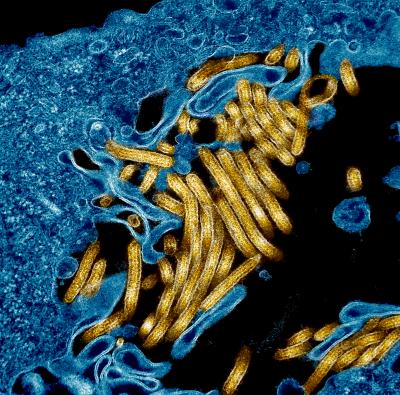
Scientists Discover New Method Ebola Virus Uses to Infect Cells

Media Type
Article
Publish or Event Date
Research Institution
Texas Biomedical Research Institute
Short Title
Scientists Discover New Method Ebola Virus Uses to Infect Cells
Content Coordinator

Scientists Discuss Prototype Pathogens for Pandemic Preparedness

NIAID Now |

NIAID Approach Highlighted in New Journal Supplement

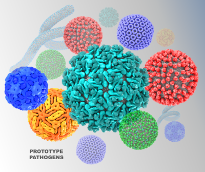
A special Oct. 19 supplement to the Journal of Infectious Diseases contains nine articles intended as a summary of a National Institute of Allergy and Infectious Diseases (NIAID)-hosted pandemic preparedness workshop that featured scientific experts on viral families of pandemic concern. Sponsored by NIAID, the supplement features articles on 10 viral families with high pandemic potential known to infect people. Concluding the supplement is a commentary from NIAID staff on the “road ahead.”

Many of the viruses in these 10 families have no vaccines or treatments licensed or in advanced development for use in people. Rather than facing the enormous task of developing medical countermeasures for individual viruses, one strategy is to use the “prototype pathogen” approach – which was shown to be successful with the rapid development of vaccines during the SARS-CoV-2 pandemic. This approach characterizes “representative” viruses within viral families so that knowledge gained, including medical countermeasures strategies, can be quickly adapted to other viruses in the same family.

The NIAID workshop on pandemic preparedness had several goals, including to describe the prototype pathogen approach, select prototype pathogens for future study, and identify knowledge gaps within the selected viral families. Prototype viruses being considered for study within the 10 families of pandemic concern are listed below. The ranges of these prototype viruses span the globe.

  • Arenaviridae: These viruses are capable of spillover from animals to people and can lead to severe viral hemorrhagic fevers. Lassa virus and Junín virus were selected as prototypes.
  • Bunyavirales, includes the Hantaviridae, Nairoviridae, Peribunyaviridae and Phenuivirdae families, among others. Viruses in this family are spread by several different arthropods (mosquitoes, ticks, midges) or rodents and can cause mild to severe symptoms and death.
    • Phenuivirdae prototypes are Rift Valley fever virus, severe fever with thrombocytopenia syndrome virus (SFTSV), Toscana virus, and Punta Toro virus.
    • Nairoviridae prototypes are Crimean-Congo hemorrhagic fever virus and Hazara virus.
    • Hantaviridae prototypes are Hantaan virus, Sin Nombre virus, and Andes virus.
    • Peribunyaviridae prototypes are La Crosse virus, Oropouche virus, and Cache Valley virus.
  • Paramyxoviridae: This family includes highly transmissible viruses that are well known (measles, mumps) and more recently emerged (Nipah virus). Viruses proposed as prototypes are Cedar virus, canine distemper virus, human parainfluenza virus 1/3, and Menangle virus.
  • Flaviviridae: These viruses, primarily transmitted by mosquitoes and ticks, are responsible for hundreds of millions of human infections worldwide each year. Viruses proposed as prototypes are West Nile virus, dengue serotype 2 virus, and tick-borne encephalitis virus.
  • Togaviridae: Most of these viruses are spread by mosquitoes and cause disease in animals that then can spillover to people. Viruses proposed as prototypes are Chikungunya virus and Venezuelan equine encephalitis virus.
  • Picornaviridae: This family includes common human viruses such as polio and hepatitis A, but new technology has led scientists to recently discover more than 300 new viruses. The four selected prototypes are enteroviruses A71 and D68, human rhinovirus C virus, and echovirus 29.
  • Filoviridae: Filoviruses can cause severe hemorrhagic fever in people and have been causative agents of recent outbreaks. Ebola virus is the prototype virus.

Experts with careers built on knowledge of each virus family are leading research teams across the U.S., studying how viruses infect cells, which models of disease most closely mimic human disease, and how to use new technology when designing vaccines and treatments. NIAID leaders are anticipating that the prototype approach will create “opportunities for investigators from multiple fields or with specialized technical expertise to collaborate in new ways.”

Reference: Pandemic Preparedness at NIAID: Prototype Pathogen Approach to Accelerate Medical Countermeasures—Vaccines and Monoclonal Antibodies. Journal of Infectious Diseases (2023).

Contact Information

Contact the NIAID Media Team.

301-402-1663
niaidnews@niaid.nih.gov

Search NIAID Blog

Amarasinghe Awarded Grant for Ebola Virus Research

Media Type
Article
Publish or Event Date
Research Institution
Washington University School of Medicine in St. Louis
Short Title
Amarasinghe Awarded Grant for Ebola Virus Research
Content Coordinator

Tijana Ivanovic, Ph.D.

Section or Unit Name
Single Virion Biology and Biophysics Unit (SVBBU)

Highlight

Exclude from directory
Off
Section/Unit: Year Established
Section/Unit: Location
This Researcher/Clinician’s Person Page
Parent Lab/Program
Program Description

Viral outbreaks and pandemics arise when animal viruses adapt so that they can recognize and use human cells as their host. Viral persistence in human circulation is subsequently driven by rapid acquisition of mutations in viral proteins that allow evasion of the adaptive human immune response.

Recent research in the lab has shown that viral ability to recognize and invade host cells in the presence of immune system antibodies is determined by the viral particle structure. For example, the same antibodies binding to the same target viral protein may or may not neutralize viral infection depending on the overall structural organization of the virus particle displaying these targets.

We are further working to define how the structural organization of virus particles might enable entry into cells not displaying fitting receptors. Viral ability to establish new infections under suboptimal conditions is probed as a possible mechanism of adaptation to new hosts with the potential to lead to new pandemics. Complementing our research on virus cell entry is our effort to define the mechanisms of virus particle assembly and regulation of the assembly outcomes (i.e., structural features of virus particles such as shape, size, or viral protein incorporation). We have developed novel quantitative and high-throughput approaches for studying virus particle assembly, and detailed functional characterizations are underway.

Trainees learn how to think deeply as virologists and acquire a unique combination of interdisciplinary skills that together permit them to seek answers to sophisticated questions. We combine molecular virology, in vitro reconstitution, biophysics, and quantitative cell biology to answer difficult-to-tackle mechanistic questions in virology and build upon those discoveries to ultimately enable prediction and prevention of undesirable viral adaptation (e.g., drug resistance, immune escape, or pandemic adaptation). Our work spans diverse viral pathogens and/or their model systems, including but not limited to influenza virus and reovirus.

Viral cell-entry mechanisms

Viruses initiate infection by delivering the genomes from within the viral capsid or a membrane envelope to the target cell. Enveloped viruses accomplish this by fusing/merging their membrane with a cellular membrane. Membrane fusion is mediated by the viral surface glycoproteins, which undergo a series of large-scale conformational changes that allow them first to insert in the target membrane, then to bring the viral and cellular membranes together. By measuring the fusion kinetics of individual virus particles and their mutants in the presence or absence of antibodies or inhibitors, we have uncovered the detailed mechanism of membrane fusion by influenza virus.

Influenza cell-entry glycoprotein hemagglutinin (HA) is present at a high density on virus particles. Three to five HAs that insert next to each other cooperate to bring about membrane fusion. However, the steps leading to HA insertion are stochastic (noncooperative, random), so substoichiometric (nonsaturating) antibody or inhibitor amounts prevent fusion by the majority of particles.

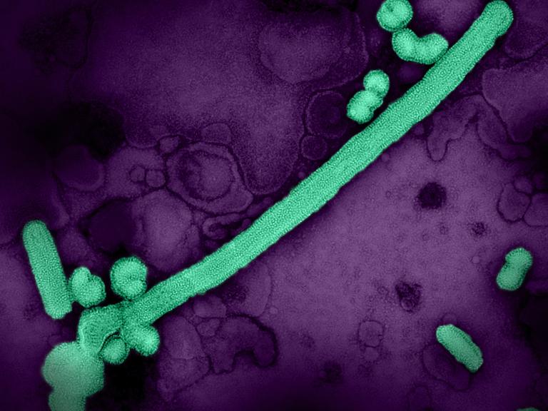
A further consequence of the mechanism of membrane fusion we uncovered is that very large particles are virtually impossible to inhibit. Tens of micrometers-long viral filaments require more than 90% of HAs to be inactivated for any reduction in the fusion yield. This insight led to a model where the pre-existing diversity in virus particle size in pleomorphic (mixed-shape) viruses enables viral adaptation to cell-entry pressure in a way that does not require an initiating genetic change.

We are asking:

  1. How does virus particle shape influence viral adaptation to cell-entry pressure?
  2. What is the mechanism of membrane fusion by other pleomorphic viruses (e.g., Ebola virus)?
  3. What role do other viral structural features (e.g., glycoprotein density) play in the mechanism of membrane fusion?
  4. What role do the interactions between viral glycoproteins and cellular receptors play in membrane fusion?
Composite of three images regarding SVBBU entry mechanisms

Left: Electron micrograph revealing pleomorphic influenza A virions. A filamentous and a spherical virion are indicated. Pleomorphic virus structure is employed by other circulating (e.g., measles and RSV) or emerging (e.g., highly pathogenic avian influenza, Ebola, Nipah, and Hendra viruses) devastating viral pathogens. Middle: Home-built TIRF microscope for single-virion and single-molecule imaging. As our research evolves, we update the design of our microscopes to meet the changing experimental needs. Right: Longest filamentous virions are effectively resistant to inhibition by HA-targeting antibodies.

Credit: NIAID

Viral assembly mechanisms

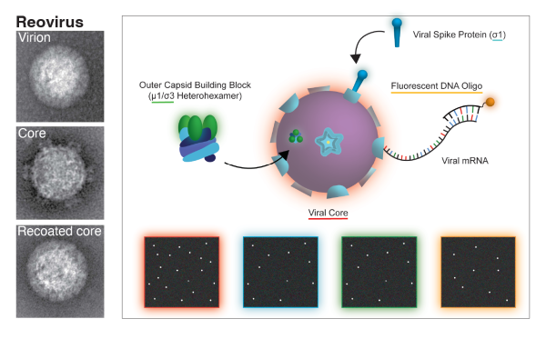
We have established a total internal reflection fluorescence (TIRF)-based single-particle platform for quantitative real-time imaging of reovirus assembly, disassembly, and transcription.

Reovirus virion consists of two concentric icosahedral protein shells. The inner, core particle encloses the genome and serves as the transcriptional unit in the infected cell cytoplasm. The outer capsid delivers the core across the cellular membrane to initiate infection. Disassembly of the outer capsid activates cores for transcription, and its assembly around cores represses transcription.

In our experiment, fluorescently labeled cores are immobilized on microscope coverslips. Fluorescently tagged oligonucleotides detect nascent viral transcripts. Fluorescently tagged outer-capsid components assemble around immobilized cores and repress transcription. We are working toward a quantitative model of reovirus outer-capsid assembly and transcriptional regulation.

In addition to studying the assembly of regularly shaped, icosahedral virions, we are interested in mechanisms of assembly of irregularly shaped pleomorphic viruses. For this, we have developed a novel, high-throughput assay enabling quantification of virus particle size distributions directly in the medium around infected cells.

We are asking:

  1. What is the mechanism of pleomorphic virion assembly?
  2. How is the shape of pleomorphic virions regulated?
  3. What are the viral and cellular determinants of viral assembly outcomes?
  4. How do viruses with segmented genomes ensure efficient packaging of the full genome complement?
Black and white photos next to a model image of SVBBU assembly mechanisms

Left: Electron micrograph of Reovirus virions, cores, and recoated cores in vitro. Reovirus virions display a regular, icosahedral particle structure. Recoated cores are indistinguishable from virions in structure and function. Right: Single particle platform for study of reovirus assembly, disassembly, and transcription. Schematic of the experiment design and illustration of what transcription/assembly might look like under the microscope. Red square: dots correspond to viral cores (all particles). Blue and green squares: cores that have incorporated the outer-capsid proteins sigma1 (blue) and mu1/sigma3 (green). Orange square: actively transcribing cores.

Credit: NIAID
Selected Publications

Partlow EA, Jaeggi-Wong A, Planitzer SD, Berg N, Li Z, Ivanovic T. Influenza A Virus Infections Sense Host Membrane Tension to Dynamically Tune Assembly. bioRxiv [Preprint], 2023 Aug 28:2023.08.28.555166.

Li Z, Li T, Liu M, Ivanovic T. Hemagglutinin Stability Determines Influenza A Virus Susceptibility to a Broad-Spectrum Fusion Inhibitor Arbidol. ACS Infec Dis. 2022 Aug 12;8(8):1543-1552. 

Li T, Li Z, Deans EE, Mittler E, Liu M, Chandran K, Ivanovic T. The shape of pleomorphic virions determines resistance to cell-entry pressure. Nat Microbiol. 2021 May;6(5):617-629. 

Ivanovic T, Harrison SC. Distinct functional determinants of influenza hemagglutinin-mediated membrane fusion. Elife. 2015 Nov 27;4:e11009. 

Ivanovic T, Choi JL, Whelan SP, van Oijen AM, Harrison SC. Influenza-virus membrane fusion by cooperative fold-back of stochastically induced hemagglutinin intermediates. Elife. 2013 Feb 19;2:e00333. 

Major Areas of Research
  • Viral cell-entry and assembly mechanisms
  • Virus particle shape regulation in pleomorphic viruses
  • Structure/function relationship for uniformly shaped, icosahedral virus particles
  • Viral adaptation – crosstalk between phenotypic and genetic diversity
  • Novel antivirals
Research Group Page

Tijana Ivanovic, Ph.D.

Education:

Postdoctoral Fellowship, 2015, Harvard University

Ph.D., Virology, 2008, Harvard University

B.S., Microbiology and Molecular Genetics, 1999, University of California, Los Angeles

Photo of Tijana Ivanovic, Ph.D.

National and Regional Biocontainment Research Facilities

The National Biocontainment Laboratories (NBLs) and Regional Biocontainment Laboratories (RBLs) provide BSL4/3/2 and BSL3/2 biocontainment facilities, respectively, for research on biodefense and emerging infectious disease agents.