Benjamin Crews is a postbac in the RML Microscopy Unit of the Research Technologies Branch (RTB) located at Rocky Mountain Laboratories in Hamilton, Montana, where he works under the supervision of Elizabeth Fischer, M.A. Read about Ben’s unique experience as a postbac in RTB.
I joined the National Institutes of Health (NIH) as a postbac fellow following undergrad and knew that I wanted to conduct research for the next two years. With such broad interests, joining the RTB was the best decision I could have made. The RTB specializes in employing cutting-edge research technologies to help NIAID investigators solve problems. RTB encompasses an array of specialties including genomics, flow cytometry, protein chemistry, visual medical arts, and more.
I am a part of the Electron Microscopy Unit headed by Beth Fischer, M.A. based at Rocky Mountain Laboratories in Hamilton, Montana. Our unit specializes in the imaging of biologic samples, specifically related to infectious diseases. NIAID researchers reach out to our group for imaging expertise, and we help determine what type of imaging best addresses their questions before processing and imaging their samples. These collaborations often involve a lot of back and forth, which allows us to learn about the experiments as the projects move forward. For a recent graduate interested in learning about many research topics, this has been the perfect experience. Our group works with a huge array of organisms and has a broad range of technologies with which we can investigate them. Whether we are using cryo-transmission electron microscopy (TEM) to investigate prion structure or conventional scanning electron microscopy (SEM) to probe the inside of the mosquito digestive tract, there is always something new and interesting to learn. This diversity of projects I get the opportunity to work on has been the most atypical aspect of my postbac experience thus far.
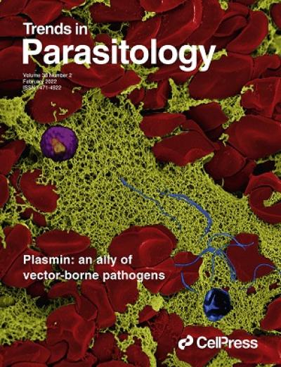
Even though we are high in the Rockies, we collaborate with researchers across the country. My most recent project was in collaboration with Zarna Pala, Ph.D., a postdoc fellow in the Molecular Parasitology and Entomology Unit in the Laboratory of Malaria and Vector Research located on the Twinbrook campus in Rockville, Maryland. Dr. Pala researches malaria parasites and was interested in how these parasites interact with fibrin in the blood. To help answer this question, she reached out to us with a request to image the malaria-infected blood bolus inside the mosquito midgut. I was assigned to this project and met virtually with both Dr. Pala and her supervisor Dr. Vega-Rodriguez to discuss their project. I received the mosquito samples in the mail from Maryland and I prepped and imaged them via SEM. Though we have not fully discovered how malaria interacts with fibrin in blood clots, Dr. Pala’s research in tandem with RTB expertise has started to uncover new and unique insights into malarial pathogenesis. Dr. Pala recently authored Beyond Cuts and Scrapes: plasmin in malaria and other vector-borne diseases published in Trends in Parasitology, which featured an image from our collaboration as the issue cover.
I plan to spend two years at the RTB while applying to medical school. Thus far, my time here has been an amazing exposure to the world of infectious disease and has offered more breadth than I could have hoped for. I know that in my future medical career, when I come across diseases I have studied here, I will be able to think of them in different and nuanced ways thanks to the special opportunities that an RTB postbac has given me.


