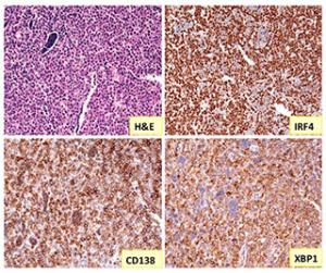
Immunohistochemical analyses of a plasmacytic plasmacytoma, which show IRF 4 expressed in the cell nucleus, CD138 on cell membrane, and XBP1 in cytosols
The Pathology Core offers up-to-date expert support to NIAID intramural investigators by providing any and all research pathology-related services. These duties include preparation, diagnosis, and reporting of research pathology samples. In addition, the core provides collaborative advice in experimental design, performance, and interpretation of results, as well as assistance in manuscript preparation involving studies using laboratory animals, all aimed at validation of our animal pathology studies.
Collaborations with the LIG Pathology Core are available. Contact Dr. Chen-Feng Qi, Chief of Pathology Core. As a senior pathology research specialist with 20 years of human and research pathology experience, Dr Qi is leading the research Pathology Core to provide immediate, relevant, and useful services to all our staff members.

Immunohistochemical analyses of a plasmacytic plasmacytoma, which show IRF 4 expressed in the cell nucleus, CD138 on cell membrane, and XBP1 in cytosols.
Support Offerings
- Web page to apprise researchers of available services and contact information
- Organ and tissue examination; interpretation and diagnoses of surgical, anatomical, and cytological research pathology samples including bone, bone marrow, blood, urine, fluid smears
Note: Tissues must be fixed by requestor. - Examination, reporting, and consultation on all mouse models from eye to skin, from urology to neurology, from cardiovascular to reproductive system, and more
- Consultation and evaluation with all histopathological specimens and scoring of immunostained slide tissue for specific cell binding with antibodies
- Dual view microscope consultation
- Written pathology reports
- Hematopathology-related experimental consultation
- Cytospin of live cell culture and cell fractionation from cell culture and fluids, including samples with H&E and Giemsa staining
- Photomicrographic reports
- Consultation on experimental design, conduct, and manuscript preparation
- Instruction and consultation for immunohistochemical studies, including antibody markers for distinguishing specific antigens and cell tissue locations; full range of specimen examination: immunofluorescence staining, confocal microscopy, specific antibody markers, tumor markers, and receptor analyses with paraffin-embedded or frozen sections
Advisory Committee
- Dr. Zheng-Ping Zhuang, M.D., Ph.D., a board-certified pathologist, director of Developmental Molecular Diagnostic Unit, National Cancer Institute Laboratory of Pathology
- Dr. Jerrold Ward, D.V.M., Ph.D., a board-certified veterinary pathologist, former chief of the Infectious Disease Pathogenesis Section, NIAID Comparative Medicine Branch
Image Gallery

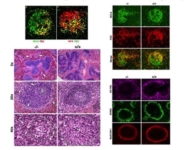
Differential expression of IRF8 in subsets of macrophages and dendritic cells.

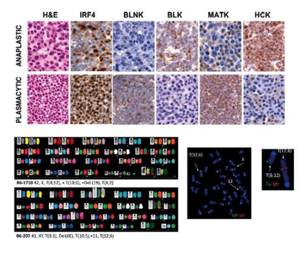
Anaplastic plasmacytomas: relationships to normal memory B cells and plasma cell neoplasms of immunodeficient and autoimmune mice

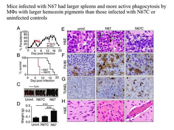
Strain-specific innate immune signaling pathways determine malarial parasitemia dynamics and host mortality

