Structural Immunology Section
Peter Sun, Ph.D.
Chief, Structural Immunology Section
Acting Chief, Laboratory of Immunogenetics
Major Areas of Research
- HIV pathogenesis and infection-induced viral release
- Structural biology of T cell receptors
- Inhibition of coronavirus release and mitigate COVID-19 pathogenesis
SIS has created a number of software programs available to all researchers. Access the SIS software programs.
Program Description
HIV pathogenesis and infection-induced viral release
Sialic-acid Ig-like binding lectins (SIGLECs) are a family of adhesion and signaling receptors that specifically recognize sialylated carbohydrate moieties. The potential involvement of members of SIGLEC family receptors in HIV pathogenesis remain to be determined. To understand the molecular basis for the sialic acid-dependent adhesion implemented by SIGLECs, and to get an insight into receptor specificity, structural studies have been carried out using two Ig-like N-terminal domains of SIGLEC-5. X-ray structure solution using molecular replacement with phased translation function uncovered unparalleled features not seen in other one-domain structures of related SIGLECs, including unusual conformation of variable loop C-C of the ligand-binding domain and a unique interdomain disulfide bond. To get an insight into receptor specificity, the group has crystallized several receptor-ligand complexes using sialylated oligosaccharides commonly found at cell surfaces and in the extracellular milieu. In combination with binding assays, these structural studies have given us an insight into what governs ligand recognition and receptor specificity in SIGLEC family of lectins.
In addition to SIGLEC receptors, the lab recently began to investigate the potential interaction between C-type lectin receptor, CD62L, and HIV-1 envelope protein, gp120. CD4 and chemokine receptors mediate HIV-1 attachment and entry. They are, however, insufficient to explain the preferential viral infection of central memory T cells. The lab identified L-selectin (CD62L) as a viral adhesion receptor on CD4 T cells. The binding of viral envelope glycans to L-selectin facilitates HIV entry and infection. L-selectin expression on central memory CD4 T cells supports their preferential infection by HIV. Upon infection, the virus downregulates L-selectin expression through shedding, resulting in an apparent loss of central memory CD4+ T cells. Infected effector memory CD4+ T cells, however, remain competent in cytokine production. Surprisingly, inhibition of L-selectin shedding markedly reduces HIV-1 infection and suppresses viral release, suggesting that selectin shedding is required for the viral release. These findings highlight a critical role for cell surface sheddases in HIV-1 release and reveal new antiretroviral strategies based on small molecular inhibitors targeted at metalloproteinases for viral release.
Currently utilized antiretroviral therapies are insufficient to eliminate HIV infection and there are no compounds targeting HIV-1 release. We showed that HIV-1 release is not spontaneous, but rather requires caspase-induced phosphatidylserine exchange to facilitate the shedding of the viral adhesion receptor L-selectin/CD62L. All three classes of caspases are activated in HIV infected cells with their activation correlated with the viral infection. Blocking of caspase activation dramatically suppressed HIV-1 infection of primary CD4+ T cells and inhibited the viral release. Budding virions in the presence of a pan-caspase inhibitor become tethered through CD62L, and exhibit reduced structural fitness. Further, HIV accessory gene nef is primarily responsible for inducing caspase activation for viral release. Importantly, caspase inhibition suppressed viral release from patient derived CD4+ T cells, from both chronically infected individuals and from aviremic viral reservoirs. These results establish a new paradigm with caspase activation critical for the release phase of HIV life cycle. It suggests a new antiviral strategy targeted at viral reservoir release by suppressing rather than stimulating apoptotic pathways.
Structural biology of T cell receptors
T cells are developed in thymus to become major histocompatibility complex (MHC)-restricted. It is generally accepted that MHC binding affinity drives T cell maturation. However, it is not clear what structural constraints imposed by MHC to shape a developing T cell receptor repertoire. In collaboration with Dr. Alfred Singer's group at the National Cancer Institute (NCI), the lab investigated the determinants for T cell receptor specificity against MHC ligands. The central feature of T cell biology is that T cells selected from the thymus utilize their antigen receptors (TCR) to specifically recognize antigenic peptides presented by the MHC, a characteristic referred to as MHC-restriction. However, the mechanisms leading to MHC-restriction of T cells are not fully understood. Previous structural studies of TCR and peptide/MHC complexes suggested that MHC-restriction is intrinsic to TCR structures as a result of germline-encoded CDR1 and CDR2 loops that have evolved to specifically promote contacts with MHC. In contrast, emerging evidence has shown that MHC-restriction is imposed by thymic selection in that coreceptor-independent TCR signaling in the thymus permits selection of TCRs that recognize MHC-independent ligands. Further deep sequencing analysis revealed that the overall germline V gene usage was similar in the peripheral TCR repertoire of both wild type B6 and Quad_KO mice. Nevertheless, individual TCR clones were selected at remarkably different frequencies in the presence or absence of MHC, further demonstrating the ligand specificity of TCRs was imposed by the thymic selection. The comparisons of TCR sequences and their frequencies between MHC-restricted and independent repertoires enabled the group to delineate any amino acid sequence preferences both in germline-encoded CDR1 and 2, and in non-germline derived CDR3 regions specific for MHC-recognition. In an attempt to characterize the usage of both germline and non-germline regions of TCR in response to MHC, the lab carried out RNA-seq based deep sequencing on TCR chains from both MHC-independent and MHC-restricted animals. To address the impact of thymic selection to TCR repertoires, the lab also sequenced TCR repertoire from pre-selection double negative (DN) B6 thymocytes. These comparative repertoire analyses of polyclonal TCRs from different mouse strains revealed that MHC-restricted, but not independent repertoires, share greater number of public TCR sequences. They selectively enrich CDR3 sequences containing smaller, hydrogen-bonding amino acids but disfavor larger, hydrophobic amino acids, and exclude cysteine residues in their MHC-binding site. In contrast, MHC-independent TCRs exhibited antibody-like CDR3 amino acid compositions. The selective preference of residue composition in CDR3, but not in germline-encoded CDR1 and 2, demonstrates that MHC specificity is not intrinsic to germline-encoded TCR sequences, but results from ligand-specific selections. In addition to comparative repertoire analyses, the lab also reported the first crystal structures of MHC-independent TCRs (A11 and B12A) and both recognized mPVR as their activation and selection ligand. B12A and A11 closely resemble the conventional MHC-restricted TCRs. The structural comparison of these MHC-independent TCRs with those of MHC-restricted ones illustrated the lack of structural changes in CDR1 and 2, demonstrating that the structures of germline V genes could not pre-determine TCR ligand specificity. Moreover, B12A and A11 TCRs recognized different epitopes on mPVR, reminiscent of antibody-antigen recognition. In summary, the results suggest that within the pre-selected repertoire, TCRs are capable of recognizing a huge diversity of ligand structures and highlight the role of thymic selection in determining the TCR specificities.
The comparison of TCR amino acid sequences of various pre-selection repertoires with those of mature repertoires from multiple MHC-specific strains, as well as MHC-independent (MHCi) animals, showed that MHC-specific thymic selection affected only non-germline encoded CDR3, restricting both their length and usage of specific amino acids, but did not affect TCR V-gene usage, nor V-J pairing. Violation of these constraints may result in T cells to fail positive selection from unfavorable interactions with MHC or undergo clonal deletion due to self-reactivity. In addition, the lab’s crystallographic structure studies on two MHCi TCR showed the receptor achieved MHC-independence without significant conformational change but rather through increasing the length of CDR3, which preclude the structural docking of MHC ligand.
Adoptive T cell immunotherapy (ACT) treats cancer by the injection of large numbers of autologous tumor specific T cells. While promising, ACT is far from effective and understanding of the mechanism is missing. One successful treatment to metastatic colorectal cancer where the tumor carried the common oncogenic KRAS-G12D mutation depended on T cell recognition of peptides derived from mutant the G12D neoantigen presented by MHC on tumors. Several TCRs specific for this mutation were identified to be restricted by HLA-C*08:02 recognizing two G12D peptide epitopes, a 9mer (GADGVGKSA) and a 10mer (GADGVGKSAL). The lab solved the high resolution crystal structures of HLA-C*08:02 presenting the 9mer and 10mer peptides alone and in complex with their cognate T cell receptors (TCRs). The TCR free HLA-C structures demonstrated a bulge in the 10mer peptide compared to the 9mer, peaking at K7. In the TCR:HLA-C-10mer structure, this bulge in the peptide shifted from K7 to V5 and is accommodated through hydrophobic contacts with the CDR3 chain, while the main peptide contact is through a Y97 in the CDR3. This conformation dependent recognition of the 10mer provides a structural explanation for why the 10mer specific TCR is unable to recognize the highly similar 9mer peptide. The TCR:HLA-C-9mer structure revealed that the 9mer peptide was recognized primarily through an E51 and Y50 of the CDR2, while the CDR3 did not contribute to peptide recognition and interacted with the HLA-C heavy chain. The CDR3 chain through Q98 contacted both the peptide at position D3 and HLA-C at R156. Additionally, the lab’s structures demonstrated that both epitopes are similarly presented by HLA-C*08:02 through a direct contribution of the G12D mutation, which forms a salt bridge between peptide D3 and R156 of HLA-C*08:02. This interaction is essential for antigen presentation of the G12D KRAS peptides. Since the wild type KRAS peptides cannot be presented by HLA-C*08:02, they are not recognized by these TCRs. Collectively, the lab’s data provides a structural basis for effective ACT to mutant KRAS G12D and offers a blueprint for rational design of peptide vaccines and TCR based immunotherapies. Further, the lab’s biochemical studies on the affinities of these TCR binding to their HLA ligands showed that all 9mer recognizing TCRs exhibited high affinities (10-100 nM), and their affinities appear to correlate with their persistence in patients during ACT therapy. In conclusion, the success of T cell transfer immunotherapy depends on the use of oligoclonal high affinity tumor specific T cells with variant peptide-HLA affinities to ensure both tumor killing and therapeutic persistence.
Inhibition of coronavirus release and mitigate COVID-19 pathogenesis
The ongoing pandemic caused by SARS-CoV-2 highlights the urgent need to develop therapeutics that can potentially lessen the clinical severity of COVID-19. The recently released sequence of coronavirus shows its spike protein being highly glycosylated with over 15 glycan sites. The glycosylation content is similar to that of HIV envelope gp120. The lab has recently shown that, in the case of HIV, the viral envelope glycan promotes viral entrance but hinders viral release. To facilitate the viral release, HIV activates host metalloproteinases to shed cell surface lectin receptors and inhibition of metalloproteinase activation suppressed HIV release (Kononchik et al, Nat Comm 2018). Inhibition of viral release is a proven effective antiviral strategy with Tamiflu being an FDA approved drug targeted at influenza virus release through inhibition of neuraminidase cleavage of sialic acid. The high degree of glycosylation on coronavirus spike protein suggests it has similar function to HIV gp120 glycan. The lab proposes to investigate the potential to inhibit SARS-CoV-2 coronavirus release through mechanisms that may be similar to HIV.
To characterize the SARS-CoV-2 spike protein interaction with lectin receptors, the lab has expressed recombinant SARS-CoV-2 spike prefusion trimer using 293F freestyle expression system. Given the nanomolar high affinity of the spike protein binding to human ACE2, the entry receptor for SARS-CoV-2, and the physiological importance of ACE2 in balancing blood pressure, the group investigated if this interaction would affect the enzymatic activity of ACE2. Surprisingly, SARS-CoV-2 trimeric spike protein increased ACE2 proteolytic activity 3-10-fold when fluorogenic caspase-1 substrate and Bradykinin-analog peptides were used to characterize ACE2 activity. In addition, the enhancement was mediated by ACE2 binding of RBD domain of SARS-CoV-2 spike. These results highlighted the altered activity of ACE2 during SARS-CoV-2 infection and would shed new lights on the pathogenesis of COVID-19 and its complications for better treatments (Lu and Sun JBC 2020).
Biography
Education
Ph.D., Molecular Biology Institute, University of Oregon
Dr. Sun obtained his Ph.D. from the Molecular Biology Institute, University of Oregon, for the study of structure and thermostability of phage T4 lysozyme using X-ray crystallography. He then joined the National Institute of Diabetes and Digestive and Kidney Diseases for his postdoctoral training in 1991, focusing on the structure and function of cytokines. In particular, he determined the crystal structure of a human transforming growth factor, TGF-beta 2. He joined NIAID in 1994.
Selected Publications
- Erickson R, Huang C, Allen C, Ireland J, Roth G, Zou Z, Lu J, Lafont BAP, Garza NL, Brumbaugh B, Zhao M, Suzuki M, Olano L, Brzostowski J, Fischer ER, Twigg HL 3rd, Johnson RF, Sun PD. SARS-CoV-2 infection of human lung epithelial cells induces TMPRSS-mediated acute fibrin deposition. Nat Commun. 2023 Oct 11;14(1):6380.
- Segura J, Ireland J, Zou Z, Roth G, Buchwald J, Shen TJ, Fischer E, Moir S, Chun TW, Sun PD. HIV-1 release requires Nef-induced caspase activation. PLoS One. 2023 Feb 13;18(2):e0281087.
- Sim MJW, Stotz Z, Lu J, Brennan P, Long EO, Sun PD. T cells discriminate between groups C1 and C2 HLA-C. Elife. 2022 May 19;11:e75670.
- Sim MJW, Sun PD. T Cell Recognition of Tumor Neoantigens and Insights Into T Cell Immunotherapy. Front Immunol. 2022 Feb 10;13:833017.
Research Group

From left: David Myers, Peter Sun, Joanna Ireland, Elizaveta Lyapina, Chang Huang, Stephen Trampe, Ted Zou, Gwynne Roth, Jinghua Lu.
Tools and Equipment
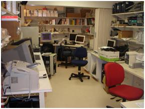
Computer room
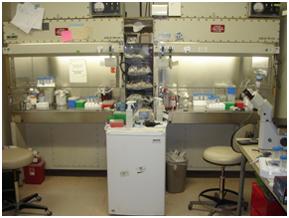
Chemical and biological hoods
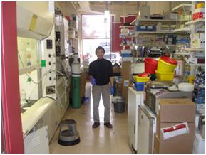
Laboratory space
Sub-zero freezers
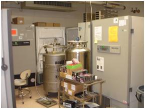
X-ray Equipment
- 2 Rigaku X-ray generators
- 1 Raxis IV imaging plate detector
- 1 Raxis IV++ imaging plate detector
- 2 LN2 cryogenic systems
Laboratory Equipment
- Gilson HPLC
- Biocad 700E
- Pharmacia FPLC
- Bioflo 3000 Bioreactor
- Microscopes
Computer Equipment
- 2 SGI O2s running IRIX3
- 8 PCs running Red Hat Enterprise Linux 5/6
- 1 Tektronix Phaser 440 printer
- 3 HP LaserJet printers
Crystallosgraphic Software
- CCP4
- Phaser
- Phenix
- Solve/Resolve
- Pymol
- CCP4MG
- XtalView
- Coot
- HKL2000
- Arp Warp
- Doc
- Dtrek
- Shelx
- Sharp
- SnB
- O
- Phases
- CNS
- APBS
- TNT